Search
« Previous |
1 - 10 of 359
|
Next »
Search Results
Select an image to start the slideshow

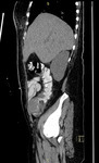
CT (abdomen), Abdominal Aortic Aneurysm
1 of 10

X-ray (chest), AP Lat - Pleural Effusion
2 of 10
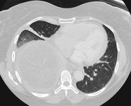
CT (chest), Pleural Effusion
3 of 10
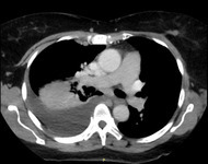
CT (chest), Pleural Effusion
4 of 10
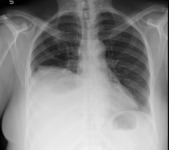
X-ray (chest), AP Lat - Pleural Effusion
5 of 10
CT (abdomen, pelvis), Sigmoid Colon Cancer
6 of 10
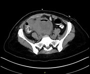
CT (abdomen, pelvis), Sigmoid Colon Cancer
7 of 10
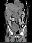
CT (abdomen, pelvis), Sigmoid Colon Cancer
8 of 10
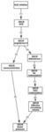
Concept diagram: Management of hypertension with ACE inhibitor
9 of 10
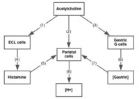
Concept diagram: Management of excess gastric acid
10 of 10
