Search

You searched for: Creator Chris DeMauro, MD Remove constraint Creator: Chris DeMauro, MD Keyword Diagnosis Remove constraint Keyword: Diagnosis Subject Respiratory Tract Diseases Remove constraint Subject: Respiratory Tract Diseases

Search Results

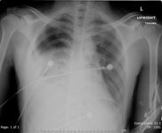
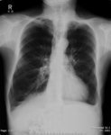
  1. X-ray (chest), AP, Post-gunshot Wound, With and Without Chest Tube

    show more
    Description: Adult male status post-gunshot wound There is a right hemothorax with a right apical pneumothorax. There is airspace disease at the right lung base. There is a small amount of subcutaneous emphysema overlying the lateral aspect of the right hemithorax. The left lung is well expanded and clear. The visualized portions of the cardiac, hilar, and mediastinal silhouettes are unremarkable. There is also a comminuted fracture of the left humerus.
    Keywords: X-Ray Radiology, Diagnostic, Roentgenography, Radiography, Diagnostic X-Ray Radiology, X-Ray, Diagnostic, postgunshot, Radiology, Diagnostic X-Ray, Diagnosis, Diagnostic X-Ray, comminuted fracture
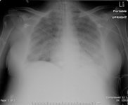
  2. X-ray (chest), PA, Adult Respiratory Distress Syndrome, ARDS, Adult Female

    show more
    Description: Chest: The endotracheal tube is unchanged in position. There bilateral diffuse pulmonary opacities. There is no pleural effusion. The cardiac silhouette is obscured. Impression: Progressive diffuse airspace disease compatible with ARDS.
    Keywords: X-Ray Radiology, Diagnostic, X-Ray, Diagnostic, Diagnostic X-Ray, ARDS, Human, Diagnosis, Roentgenography, Radiography, Shock Lung, Radiology, Diagnostic X-Ray, Acute Respiratory Distress Syndrome, Diagnostic X-Ray Radiology
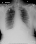
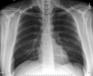
  3. X-ray (chest), AP, Pneumothorax with Chest Tube

    show more
    Description: Adult female, with chest tube. There is a large right-sided pneumothorax. The left lung is well aerated and clear. The heart is not enlarged. The pulmonary vasculature is not congested. IMPRESSION: Large right pneumothorax.
    Keywords: Primary Spontaneous Pneumothorax, Spontaneous Pneumothorax, Diagnosis, Pressure Pneumothorax, Roentgenography, Tension Pneumothorax, chest tube, Radiology, Diagnostic X-Ray, X-Ray Radiology, Diagnostic, Diagnostic X-Ray Radiology, X-Ray, Diagnostic, Radiography, Diagnostic X-Ray, Pneumothorax
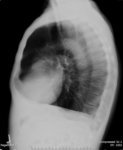
  4. X-ray (chest), Lateral, Adult male, Chronic Obstructive Pulmonary Diseaess (COPD)

    show more
    Description: The lungs are hyperinflated with apical bullous disease. There is flattening of the diaphragm on the lateral view. There is no focal infiltrate. There is no pleural effusion. The heart is mildly enlarged in size. There is no acute bony abnormality. Impression: Hyperinflation with bullous disease consistent with chronic obstructive pulmonary disease.
    Keywords: COPD, Radiography, Chronic Obstructive Pulmonary Disease, Diagnostic X-Ray Radiology, Roentgenography, X-Ray Radiology, Diagnostic, X-Ray, Diagnostic, Chronic Airflow Obstruction, Diagnosis, COAD, Chronic Obstructive Lung Disease, Radiology, Diagnostic X-Ray, Diagnostic X-Ray, Chronic Obstructive Airway Disease
  5. X-ray (chest), PA, Adult Male, Chronic Obstructive Pulmonary Diseaess (COPD)

    show more
    Description: The lungs are hyperinflated with apical bullous disease. There is flattening of the diaphragm on the lateral view. There is no focal infiltrate. There is no pleural effusion. The heart is mildly enlarged in size. There is no acute bony abnormality. Impression: Hyperinflation with bullous disease consistent with chronic obstructive pulmonary disease.
    Keywords: Chronic Obstructive Airway Disease, X-Ray Radiology, Diagnostic, COAD, COPD, Roentgenography, Chronic Obstructive Lung Disease, X-Ray, Diagnostic, Diagnostic X-Ray, Diagnosis, Chronic Obstructive Pulmonary Disease, Radiology, Diagnostic X-Ray, Radiography, Chronic Airflow Obstruction, Diagnostic X-Ray Radiology
  6. X-ray (chest), AP, Asthma with Pneumomediastinum, Adult Male

    show more
    Description: History: Asthma Frontal and lateral views of the chest were performed. Correlation is made with the CT chest also obtained on 6/3/2007. The lungs are well expanded and clear. There is extensive subcutaneous emphysema bilaterally along the neck as well as in the anterior chest. There is pneumomediastinum with air tracking along the lateral aspects of the heart. The heart is normal in size. There is no pleural effusion or pneumothorax. The visualized bones are normal. Impression: Extensive pneumomediastinum and subcutaneous emphysema in the neck as seen on the CT chest from 6/3/2007.
    Keywords: Radiology, Diagnostic X-Ray, X-Ray, Diagnostic, Diagnostic X-Ray Radiology, X-Ray Radiology, Diagnostic, Radiography, Diagnostic X-Ray, Respiratory Tract Diseases, Diagnosis, Bronchial Diseases, Roentgenography, Asthma
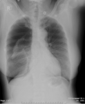
  7. X-ray (chest), AP, Pneumothorax without Chest Tube

    show more
    Description: Adult female, without chest tube. There is a large right-sided pneumothorax. The left lung is well aerated and clear. The heart is not enlarged. The pulmonary vasculature is not congested. IMPRESSION: Large right pneumothorax.
    Keywords: Roentgenography, Spontaneous Pneumothorax, Pneumothorax, Diagnostic X-Ray Radiology, Diagnosis, Radiology, Diagnostic X-Ray, X-Ray Radiology, Diagnostic, Diagnostic X-Ray, Tension Pneumothorax, Primary Spontaneous Pneumothorax, Radiography, Pressure Pneumothorax, X-Ray, Diagnostic
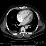
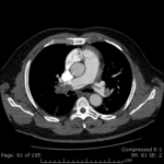
  8. CT (chest), (axial), Adult Male Pulmonary Embolism with Right Heart Strain

    show more
    Description: There are occlusive pulmonary emboli within the left and right main pulmonary arteries. The thrombus within the right main pulmonary artery extends into the right lower lobe segmental arteries. The heart is slightly enlarged with enlargement of the ventricle deviating the intraventricular septum indicating right heart strain. Additionally the main pulmonary artery measures approximately 3.2 cm in diameter indicating pulmonary arterial hypertension. Lung windows demonstrates patchy consolidation at the right lower lobe peripherally which represents infarcted lung parenchyma. There is a small right-sided pleural effusion. The left lung is clear. There is no evidence of enlarged hilar or mediastinal lymphadenopathy. The visualized portions of the upper abdominal viscera including the adrenal glands are normal. Evaluation of bone windows demonstrate no abnormality. IMPRESSION: Bilateral central pulmonary emboli with left lower lobe pneumonia infarction.
    Keywords: Pulmonary, X Ray Tomography, Computed, CAT Scan, X-Ray, Tomography, X Ray Computed, X-Ray Computerized Axial Tomography, Tomography, X-Ray Computerized, Tomography, X-Ray Computer Assisted, Tomography, X-Ray Computerized Axial, Tomography, Xray Computed, Computed Tomography, X-Ray, Tomodensitometry, Cine-CT, X-Ray Tomography, Computed, CT X Ray, Pulmonary Embolism, X-Ray Computer Assisted Tomography, Computerized Tomography, X-Ray, X Ray Computerized Tomography, Electron Beam Computed Tomography, CT Scan, X-Ray, Thromboembolism, Computerized Tomography, X Ray, Tomography, Transmission Computed, Respiratory Tract Diseases, Diagnosis, Lung Diseases, Embolism, CAT Scan, X Ray, Computed X Ray Tomography, Electron Beam Tomography, CT Scan, Axial Plane
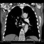
  9. CT (chest), (coronal), Adult Male Pulmonary Embolism with Right Heart Strain

    show more
    Description: There are occlusive pulmonary emboli within the left and right main pulmonary arteries. The thrombus within the right main pulmonary artery extends into the right lower lobe segmental arteries. The heart is slightly enlarged with enlargement of the ventricle deviating the intraventricular septum indicating right heart strain. Additionally the main pulmonary artery measures approximately 3.2 cm in diameter indicating pulmonary arterial hypertension. Lung windows demonstrates patchy consolidation at the right lower lobe peripherally which represents infarcted lung parenchyma. There is a small right-sided pleural effusion. The left lung is clear. There is no evidence of enlarged hilar or mediastinal lymphadenopathy. The visualized portions of the upper abdominal viscera including the adrenal glands are normal. Evaluation of bone windows demonstrate no abnormality. IMPRESSION: Bilateral central pulmonary emboli with left lower lobe pneumonia infarction.
    Keywords: Cine-CT, X Ray Tomography, Computed, Tomography, X-Ray Computerized, Pulmonary Embolism, Computerized Tomography, X-Ray, CT Scan, X-Ray, Electron Beam Computed Tomography, Computed Tomography, X-Ray, Computed X Ray Tomography, Thromboembolism, Tomography, Transmission Computed, CAT Scan, X-Ray, Tomography, X Ray Computed, X-Ray Computer Assisted Tomography, Pulmonary, Diagnosis, CAT Scan, X Ray, X-Ray Computerized Axial Tomography, Respiratory Tract Diseases, Computerized Tomography, X Ray, Lung Diseases, Tomography, Xray Computed, Tomography, X-Ray Computer Assisted, CT X Ray, Embolism, Tomography, X-Ray Computerized Axial, Electron Beam Tomography, Tomodensitometry, X Ray Computerized Tomography, X-Ray Tomography, Computed, Coronal Plane
  10. CT (chest), (axial), Adult Male, Pulmonary Embolism

    show more
    Description: There are occlusive pulmonary emboli within the left and right main pulmonary arteries. The thrombus within the right main pulmonary artery extends into the right lower lobe segmental arteries. The heart is slightly enlarged with enlargement of the ventricle deviating the intraventricular septum indicating right heart strain. Additionally the main pulmonary artery measures approximately 3.2 cm in diameter indicating pulmonary arterial hypertension. Lung windows demonstrates patchy consolidation at the right lower lobe peripherally which represents infarcted lung parenchyma. There is a small right-sided pleural effusion. The left lung is clear. There is no evidence of enlarged hilar or mediastinal lymphadenopathy. The visualized portions of the upper abdominal viscera including the adrenal glands are normal. Evaluation of bone windows demonstrate no abnormality. IMPRESSION: Bilateral central pulmonary emboli with left lower lobe pneumonia infarction.
    Keywords: Electron Beam Tomography, Thromboembolism, Cine-CT, Pulmonary Embolism, Diagnosis, Computed X Ray Tomography, Tomography, X Ray Computed, CAT Scan, X Ray, CT X Ray, Computed Tomography, X-Ray, CT Scan, X-Ray, X Ray Tomography, Computed, Computerized Tomography, X-Ray, Tomography, Xray Computed, Tomography, X-Ray Computerized Axial, Lung Diseases, X Ray Computerized Tomography, Embolism, X-Ray Computerized Axial Tomography, Electron Beam Computed Tomography, X-Ray Tomography, Computed, Tomography, X-Ray Computerized, Tomodensitometry, CAT Scan, X-Ray, Respiratory Tract Diseases, X-Ray Computer Assisted Tomography, Pulmonary, Tomography, Transmission Computed, Tomography, X-Ray Computer Assisted, Computerized Tomography, X Ray, CT Scan, Axial Plane