Search
Search Results
- Title:
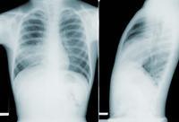
- X-ray (chest), Pneumonia RLL
- Description:
- This image depicted two chest x-rays, which revealed pathologic changes in a patient’s lung fields due to a condition known as mycoplasma pneumonia, caused by a Mycoplasma pneumoniae bacterial infection. Note on the anteroposterior (AP) view on the left, the generalized infiltrate permeating both lung fields, and consolidation in the region of the right lower lobe, as well as bilateral hilar adenopathy. In the left lateral view on the right, you can see that the consolidation occupied more of the posterior aspect of the lung fields, almost obliterating a view of the spinal column.
- Keyword:
- X-ray, Diagnositic, Lung inflammation, Pneumonitis, Pneumonia, Pulmonary inflammation, Lobar pneumonia
- Subject:
- Diagnostic Techniques and Procedures, Diagnosis
- Creator:
- Courtney Anderson
- Publisher:
- i-Human Patients, Inc.
- Copyright Holder:
- Centers for Disease Control and Prevention
- Rights:
- http://creativecommons.org/publicdomain/mark/1.0/
- Resource Type:
- Medical Imaging

- Title:
- UltrasoundOvarianCyst
- Description:
- thin-walled hypoechoic ovarian cyst
- Subject:
- Ultrasonography, Diagnostic Imaging, Diagnostic Techniques and Procedures, Diagnosis, pelvis
- Creator:
- Courtney Anderson
- Rights:
- http://creativecommons.org/licenses/by/3.0/us/
- Resource Type:
- Medical Imaging
