Search
Search Results
- Title:
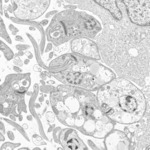
- Electron Micrograph: Leishmania braziliensis promastigotes
- Description:
- This is an electron microscopic (EM) image of Leishmania braziliensis promastigotes grown in cell culture. Note the dense kinetoplasts in the cytoplasm of these flagellated forms.
Promastigotes are not found in human tissue; this stage occurs in the mid-gut of the sand fly (genera Phlebotomus and Lutzomyia) intermediate hosts. Promastigotes are elongate, slender and measure about 10-12 µm in length. They have a large central nucleus and a kinetoplast located near the anterior end. A flagellum arises at the anterior end, that may be longer than the rest of the promastigote.
- Subject:
- Leishmaniasis, Protozoan Infections, Parasitic Diseases, Disease, Euglenozoa Infections, Pathologic Processes, Pathological Conditions, Signs and Symptoms
- Creator:
- Cynthia Goldsmith and Luciana Flannery
- Copyright Holder:
- CDC
- Rights:
- http://creativecommons.org/publicdomain/mark/1.0/
- Resource Type:
- Medical Imaging
- Title:
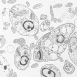
- Electron Micrograph: Leishmania major
- Description:
- This is an electron microscopic (EM) image of Leishmania major amastigotes grown in cell culture. Note the dense kinetoplasts in the cytoplasm.
Amastigotes of Leishmania are spherical to ovoid and measure 1-5 µm long by 1-2 µm wide. They possess a large nucleus, a prominent kinetoplast, and a short axoneme, the last of which is rarely visible by light microscopy. The organisms reside in macrophages of the host and can be found throughout the body.
- Subject:
- Leishmania, Disease, Parasitic Diseases, Protozoan Infections, Leishmaniasis, Trypanosomatina, Kinetoplastida, Euglenozoa, Eukaryota, Pathologic Processes, Pathological Conditions, Signs and Symptoms, Euglenozoa Infections
- Creator:
- Cynthia Goldsmith and Luciana Flannery
- Copyright Holder:
- CDC
- Rights:
- http://creativecommons.org/publicdomain/mark/1.0/
