Search
Search Results
- Title:
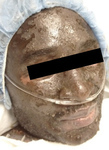
- Toxic Epidermal Necrolysis, Image 1
- Description:
- Toxic epidermal necrolysis (TEN) begins with fever, cough, and other nonspecific symptoms, and is soon followed by purplish, bloody-looking lesions on the skin and mucous membranes. These early lesions, typically found on the head, neck, and upper chest, soon merge and blister. Sheets of epidermis then begin to detach from the skin layers below. In time, the entire surface of the skin may be involved, with detachment of 100% of the epidermis.
- Keyword:
- Skin lesion, blister, Stevens-Johnson Syndrome, mucous membrane lesion
- Subject:
- Skin and Connective Tissue Diseases
- Creator:
- Metropolitan Hospital Center, Kathryn Russel, MD
- Publisher:
- Metropolitan Hospital Center
- Language:
- English
- Copyright Holder:
- Metropolitan Hospital Center
- Rights:
- http://www.i-human.com/service-agreement-print
- Resource Type:
- Photo

- Title:
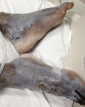
- Toxic Epidermal Necrolysis, Image 3
- Description:
- Toxic epidermal necrolysis (TEN) begins with fever, cough, and other nonspecific symptoms, and is soon followed by purplish, bloody-looking lesions on the skin and mucous membranes. These early lesions, typically found on the head, neck, and upper chest, soon merge and blister. Sheets of epidermis then begin to detach from the skin layers below. In time, the entire surface of the skin may be involved, with detachment of 100% of the epidermis.
- Keyword:
- Skin lesion, Stevens-Johnson Syndrome, mucous membrane lesion, blister
- Subject:
- Skin and Connective Tissue Diseases
- Creator:
- Metropolitan Hospital Center, Kathryn Russel, MD
- Publisher:
- Metropolitan Hospital Center
- Language:
- English
- Copyright Holder:
- Metropolitan Hospital Center
- Rights:
- http://www.i-human.com/service-agreement-print
- Resource Type:
- Photo
- Title:
- Toxic Epidermal Necrolysis, Image 2
- Description:
- Toxic epidermal necrolysis (TEN) begins with fever, cough, and other nonspecific symptoms, and is soon followed by purplish, bloody-looking lesions on the skin and mucous membranes. These early lesions, typically found on the head, neck, and upper chest, soon merge and blister. Sheets of epidermis then begin to detach from the skin layers below. In time, the entire surface of the skin may be involved, with detachment of 100% of the epidermis.
- Keyword:
- blister, mucous membrane lesion, Skin lesion, Stevens-Johnson Syndrome
- Subject:
- Skin and Connective Tissue Diseases
- Creator:
- Metropolitan Hospital Center, Kathryn Russel, MD
- Publisher:
- Metropolitan Hospital Center
- Language:
- English
- Copyright Holder:
- Metropolitan Hospital Center
- Rights:
- http://www.i-human.com/service-agreement-print
- Resource Type:
- Photo
