Search
« Previous |
51 - 60 of 110
|
Next »
Search Results
- Title:
- i-Human Case Player Tutorial - Exercises (short)
- Description:
- Case help video for exercises with abbreviated introduction.
- Subject:
- Education
- Creator:
- craig@i-human.com
- Copyright Holder:
- i-Human Patients, Inc.
- Rights:
- http://www.i-human.com/service-agreement-print
- Resource Type:
- Video

- Title:
- i-Human Case Player Tutorial - Tests & Diagnosis (short)
- Description:
- Case help video for tests with abbreviated introduction.
- Subject:
- Education
- Creator:
- craig@i-human.com
- Copyright Holder:
- i-Human Patients, Inc.
- Rights:
- http://www.i-human.com/service-agreement-print
- Resource Type:
- Video
- Title:
- i-Human Case Player Tutorial - Patient Assessment (short)
- Description:
- Case help video for assessment with abbreviated introduction.
- Subject:
- Education
- Creator:
- craig@i-human.com
- Copyright Holder:
- i-Human Patients, Inc.
- Rights:
- http://www.i-human.com/service-agreement-print
- Resource Type:
- Video
- Title:
- i-Human Case Player Tutorial - Physical Exams (short)
- Description:
- Case help video for physical exams with abbreviated introduction.
- Subject:
- Education
- Creator:
- craig@i-human.com
- Copyright Holder:
- i-Human Patients, Inc.
- Rights:
- http://www.i-human.com/service-agreement-print
- Resource Type:
- Video
- Title:
- i-Human Case Player Tutorial - Patient History (short)
- Description:
- Case help video for history taking with abbreviated introduction.
- Subject:
- Education
- Creator:
- craig@i-human.com
- Publisher:
- i-
- Copyright Holder:
- i-Human Patients, Inc.
- Rights:
- http://www.i-human.com/service-agreement-print
- Resource Type:
- Video
- Title:
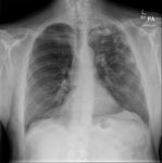
- X-ray (chest), PA, Adult Male, Tuberculosis
- Description:
- Findings:
Soft tissues of chest wall are unremarkable. Bones are intact.
Cardiomediastinal silhouette, aorta and pulmonary vasculature are normal.
Left hilar region appears elevated and there are streaky densities extending into the left upper lung zone. There is patchy density with some areas of confluence in the left upper lobe, more so in the apical region. Some cavitary areas are identified. The remainder of the lungs appear relatively clear with mild pulmonary hyperexpansion. There is left apical pleural thickening, remainder of the costophrenic angles are sharp.
Subdiaphragmatic structures are normal.
Impression: Infiltrates in left upper lobe, pleural thickening, hilar elevation, streaky densities and areas of cavitation are consistent with tuberculosis. Also consider old tuberculosis changes with superimposed pneumonia. Mild asthmatic changes also seen.
- Subject:
- Tuberculosis, Radiology, Mycobacterium Infections, Actinomycetales Infections, Gram-Positive Bacterial Infections, Bacterial Infections, Bacterial Infections and Mycoses
- Creator:
- craig@i-human.com
- Copyright Holder:
- Mammoth Hospital
- Rights:
- http://www.i-human.com/service-agreement-print
- Resource Type:
- Medical Imaging
- Title:
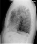
- X-ray (chest), Lateral, Adult Male, Tuberculosis
- Description:
- Findings:
Soft tissues of chest wall are unremarkable. Bones are intact.
Cardiomediastinal silhouette, aorta and pulmonary vasculature are normal.
Left hilar region appears elevated and there are streaky densities extending into the left upper lung zone. There is patchy density with some areas of confluence in the left upper lobe, more so in the apical region. Some cavitary areas are identified. The remainder of the lungs appear relatively clear with mild pulmonary hyperexpansion. There is left apical pleural thickening, remainder of the costophrenic angles are sharp.
Subdiaphragmatic structures are normal.
Impression: Infiltrates in left upper lobe, pleural thickening, hilar elevation, streaky densities and areas of cavitation are consistent with tuberculosis. Also consider old tuberculosis changes with superimposed pneumonia. Mild asthmatic changes also seen.
- Subject:
- Tuberculosis, Radiology, Mycobacterium Infections, Actinomycetales Infections, Gram-Positive Bacterial Infections, Bacterial Infections, Bacterial Infections and Mycoses
- Creator:
- craig@i-human.com
- Copyright Holder:
- Mammoth Hospital
- Rights:
- http://www.i-human.com/service-agreement-print
- Resource Type:
- Medical Imaging
- Title:
- X-ray (chest), Adult Male, Tuberculosis
- Description:
- Findings:
Soft tissues of chest wall are unremarkable. Bones are intact.
Cardiomediastinal silhouette, aorta and pulmonary vasculature are normal.
Left hilar region appears elevated and there are streaky densities extending into the left upper lung zone. There is patchy density with some areas of confluence in the left upper lobe, more so in the apical region. Some cavitary areas are identified. The remainder of the lungs appear relatively clear with mild pulmonary hyperexpansion. There is left apical pleural thickening, remainder of the costophrenic angles are sharp.
Subdiaphragmatic structures are normal.
Impression: Infiltrates in left upper lobe, pleural thickening, hilar elevation, streaky densities and areas of cavitation are consistent with tuberculosis. Also consider old tuberculosis changes with superimposed pneumonia. Mild asthmatic changes also seen.
- Keyword:
- Bacterial Infections and Mycoses, Bacterial Infections, Gram-Positive Bacterial Infections, Actinomycetales Infections, Mycobacterium Infections, Tuberculosis, Tuberculosis, Pulmonary
- Subject:
- Radiology, Tuberculosis
- Creator:
- craig@i-human.com
- Copyright Holder:
- Mammoth Hospital
- Rights:
- http://www.i-human.com/service-agreement-print
- Resource Type:
- Medical Imaging

- Title:
- i-Human Medical Image Viewer Tutorial
- Description:
- Video tutorial on the medical image viewer
- Keyword:
- MIV
- Subject:
- Delivery of Health Care, Quality of Health Care, Patient Care Management, Health Services Administration
- Creator:
- craig@i-human.com
- Copyright Holder:
- i-Human Patients, Inc
- Rights:
- All rights reserved
- Resource Type:
- Video
- Title:
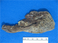
- Gross Anatomy - Lung with Interstitial pulmonary fibrosis.png
- Creator:
- craig@i-human.com
