Search
« Previous |
1 - 10 of 23
|
Next »
Search Results
- Title:
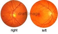
- Retinas, Normal
- Description:
- Retinas - Normal
Retina is the innermost of the three tunics of the eye, surrounding the vitreous body and continuous posteriorly with the optic nerve. The retina is composed of light-sensitive neurons arranged in three layers; the first layer is made up of rods and cones and the other two transmit impulses from the rods and cones to the optic nerve. The rods are sensitive in dim light, and the cones are sensitive in bright light and are responsible for color vision.
- Keyword:
- cones, Eye, rods, neurons, tunic
- Subject:
- Eye, Retina, Sense Organs
- Publisher:
- i-Human Patients, Inc.
- Language:
- English
- Copyright Holder:
- i-Human Patients, Inc.
- Rights:
- http://www.i-human.com/service-agreement-print
- Resource Type:
- Photo
- Identifier:
- 1646
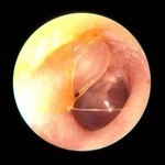
- Title:
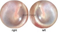
- Tympanic Membranes, Normal
- Description:
- Tympanic membranes - normal
Tympanic membrane a thin, semitransparent membrane, nearly oval in shape, that stretches across the ear canal and separates the tympanum (middle ear) from the external acoustic meatus (outer ear); called also eardrum. It is composed of fibrous tissue, covered with skin on the outside and mucous membrane on the inside. It is constructed so that it can vibrate freely with audible sound waves that travel inward from outside. The handle of the malleus of the middle ear is attached to the center of the membrane and receives the vibrations it collects, transmitting them to the other ossicles of the middle ear (the incus and stapes), which in turn transmit the vibrations to the fluid of the inner ear.
- Keyword:
- membrane, Ear, ear canal, ear drum
- Subject:
- Tympanic Membrane, Ear, Sense Organs
- Creator:
- Michael Hawke, MD
- Publisher:
- i-Human Patients, Inc.
- Language:
- English
- Copyright Holder:
- Michael Hawke, MD
- Rights:
- http://www.i-human.com/service-agreement-print
- Resource Type:
- Photo
- Identifier:
- 1645
- Title:
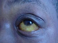
- Icteric Sclera - Jaundice
- Description:
- Icteric sclera - jaundice
- Keyword:
- Jaundice, yellow, eyes
- Subject:
- Pathological Conditions, Signs and Symptoms, Sense Organs, Sclera, Jaundice, Pathologic Processes, Eye
- Publisher:
- i-Human Patients, Inc.
- Language:
- English
- Copyright Holder:
- i-Human Patients, Inc.
- Rights:
- http://www.i-human.com/service-agreement-print
- Resource Type:
- Photo
- Identifier:
- 1635
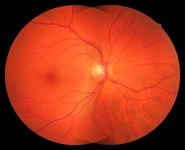
- Title:
- Normal Retina, Od Os, Funduscopic Exam
- Description:
- Normal retinas, 46 y/o female
- Keyword:
- Diagnosis, Diagnostic Techniques, Ophthalmological, eye exam, Eye
- Subject:
- Retina, Ophthalmoscopy, Eye, Diagnostic Techniques and Procedures, Sense Organs
- Creator:
- Nidek, UC Davis Ophthalmic Imaging Center
- Publisher:
- UC Davis Ophthalmic Imaging Center
- Language:
- English
- Copyright Holder:
- University of California, Davis
- Rights:
- http://www.i-human.com/service-agreement-print
- Resource Type:
- Photo
- Identifier:
- 1407
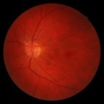
- Title:
- Normal Retina, Od Os, Funduscopic Exam
- Description:
- Normal retinas, 46 y/o female
- Keyword:
- Diagnostic Techniques, Ophthalmological, Diagnosis, Eye, eye exam
- Subject:
- Diagnostic Techniques and Procedures, Eye, Ophthalmoscopy, Retina, Sense Organs
- Creator:
- Nidek, UC Davis Ophthalmic Imaging Center
- Publisher:
- UC Davis Ophthalmic Imaging Center
- Language:
- English
- Copyright Holder:
- University of California, Davis
- Rights:
- http://www.i-human.com/service-agreement-print
- Resource Type:
- Photo
- Identifier:
- 1407
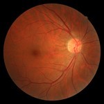
- Title:
- Normal, Funduscopic Exam
- Description:
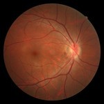
- Normal retina, 45 y/o male
- Keyword:
- eye exam, Eye, Diagnosis, Diagnostic Techniques, Ophthalmological
- Subject:
- Sense Organs, Eye, Diagnostic Techniques and Procedures, Ophthalmoscopy
- Creator:
- Nidek, UC Davis Ophthalmic Imaging Center
- Publisher:
- UC Davis Ophthalmic Imaging Center
- Language:
- English
- Copyright Holder:
- University of California, Davis
- Rights:
- http://www.i-human.com/service-agreement-print
- Resource Type:
- Photo
- Identifier:
- 1408
- Title:
- Normal, Funduscopic Exam
- Description:
- Eye, eye exam
- Keyword:
- Diagnostic Techniques, Ophthalmological, Eye, Diagnosis, eye exam
- Subject:
- Eye, Ophthalmoscopy, Diagnostic Techniques and Procedures, Sense Organs
- Creator:
- Nidek, UC Davis Ophthalmic Imaging Center
- Publisher:
- UC Davis Ophthalmic Imaging Center
- Language:
- English
- Copyright Holder:
- University of California, Davis
- Rights:
- http://www.i-human.com/service-agreement-print
- Resource Type:
- Photo
- Identifier:
- 1401
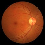
- Title:
- Eye Fundus, Normal OD
- Description:
- Normal retina 47 y/o female OD
- Keyword:
- Eye, eye exam, Diagnostic Techniques, Ophthalmological, Diagnosis
- Subject:
- Sense Organs, Diagnostic Techniques and Procedures, Ophthalmoscopy, Eye
- Creator:
- Nidek, UC Davis Ophthalmic Imaging Center
- Publisher:
- i-Human Patients, Inc.
- Language:
- English
- Copyright Holder:
- University of California, Davis
- Rights:
- http://www.i-human.com/service-agreement-print
- Resource Type:
- Photograph
- Identifier:
- 1400
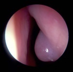
- Title:
- Partial Cast Of Tympanic Membrane, Tympanic Membrane
- Description:
- Tympanic membrane - Partial cast of tympanic membrane
Tympanic membrane is a thin, semitransparent membrane, nearly oval in shape, that stretches across the ear canal and separates the tympanum (middle ear) from the external acoustic meatus (outer ear); called also eardrum. It is composed of fibrous tissue, covered with skin on the outside and mucous membrane on the inside. It is constructed so that it can vibrate freely with audible sound waves that travel inward from outside. The handle of the malleus of the middle ear is attached to the center of the membrane and receives the vibrations it collects, transmitting them to the other ossicles of the middle ear (the incus and stapes), which in turn transmit the vibrations to the fluid of the inner ear.
- Keyword:
- membrane, ear drum, Ear, ear canal
- Subject:
- Ear, Tympanic Membrane, Sense Organs
- Creator:
- Michael Hawke, MD
- Publisher:
- i-Human Patients, Inc.
- Language:
- English
- Copyright Holder:
- Michael Hawke, MD
- Rights:
- http://www.i-human.com/service-agreement-print
- Resource Type:
- Photo
- Identifier:
- 1302
- Title:
- Anterior Nasal Mucosal Changes 2o To Chronic Nasopharyngeal Obstruction - Large Adenoids, Tympanic Membrane
- Description:
- Tympanic membrane - Anterior nasal mucosal changes 2o to chronic nasopharyngeal obstruction - large adenoids
Adenoids are one of two masses of lymphatic tissue situated on the posterior wall of the nasopharynx behind the posterior nares. During childhood these masses often swell and block the passage of air from the nasal cavity into the pharynx, preventing the child from breathing through the nose. Also called pharyngeal tonsils.
Tympanic membrane is a thin, semitransparent membrane, nearly oval in shape, that stretches across the ear canal and separates the tympanum (middle ear) from the external acoustic meatus (outer ear); called also eardrum. It is composed of fibrous tissue, covered with skin on the outside and mucous membrane on the inside. It is constructed so that it can vibrate freely with audible sound waves that travel inward from outside. The handle of the malleus of the middle ear is attached to the center of the membrane and receives the vibrations it collects, transmitting them to the other ossicles of the middle ear (the incus and stapes), which in turn transmit the vibrations to the fluid of the inner ear.
- Keyword:
- middel ear, nasopharynx, Lymphatic tissue, tonsils
- Subject:
- Sense Organs, Tympanic Membrane, Adenoids, Respiratory System, Ear
- Creator:
- Michael Hawke, MD
- Publisher:
- i-Human Patients, Inc.
- Language:
- English
- Copyright Holder:
- Michael Hawke, MD
- Rights:
- http://www.i-human.com/service-agreement-print
- Resource Type:
- Photo
- Identifier:
- 1278
