Search
« Previous |
121 - 130 of 960
|
Next »
Search Results
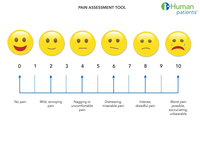
- Title:
- Pain Assessment Scale
- Description:
- Illustration of pain scale faces
- Keyword:
- history questions, history taking, Diagnosis, clinical reasoning skills, patient interview, patient history, pivotal questions
- Subject:
- Diagnostic Techniques and Procedures, Pain
- Creator:
- i-Human, Kristina
- Publisher:
- i-Human, Kristina
- Language:
- English
- Rights:
- http://www.i-human.com/service-agreement-print
- Resource Type:
- Illustration
- Identifier:
- 3243
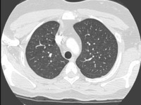
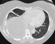
- Title:
- CT (chest), Normal
- Description:
- Normal lung
- Keyword:
- Anatomy & histology, histology, morphology, anatomy
- Subject:
- Respiratory System, Lung
- Creator:
- Jaime Drewes, MD
- Publisher:
- i-Human Patients, Inc.
- Language:
- English
- Rights:
- http://www.i-human.com/service-agreement-print
- Resource Type:
- Medical Imaging
- Identifier:
- 3236
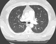
- Title:
- CT (chest), Normal
- Description:
- Normal lung
- Keyword:
- Anatomy & histology, anatomy, morphology, histology
- Subject:
- Respiratory System, Lung
- Creator:
- Jaime Drewes, MD
- Publisher:
- i-Human Patients, Inc.
- Language:
- English
- Rights:
- http://www.i-human.com/service-agreement-print
- Resource Type:
- Medical Imaging
- Identifier:
- 3236
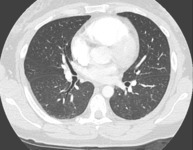
- Title:
- CT (chest), Normal
- Description:
- Normal lung
- Keyword:
- Anatomy & histology, histology, anatomy, morphology
- Subject:
- Respiratory System, Lung
- Creator:
- Jaime Drewes, MD
- Publisher:
- i-Human Patients, Inc.
- Language:
- English
- Rights:
- http://www.i-human.com/service-agreement-print
- Identifier:
- 3236

- Title:
- CT (chest), Normal
- Description:
- Normal lung
- Keyword:
- Anatomy & histology, morphology, histology, anatomy
- Subject:
- Respiratory System, Lung
- Creator:
- Jaime Drewes, MD
- Publisher:
- i-Human Patients, Inc.
- Language:
- English
- Rights:
- http://www.i-human.com/service-agreement-print
- Resource Type:
- Medical Imaging
- Identifier:
- 3236

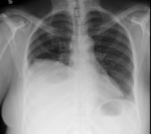
- Title:
- X-ray (chest), AP Lat - Pleural Effusion
- Description:
- There is a large opacity in the right lung base consistent with a large right pleural effusion. This was not present at the time of the comparison radiograph.
The radiographic appearance of the chest is otherwise unremarkable. The heart is normal in size. The aerated portions of both lungs appear normal. The left hemidiaphragm is well-defined, as is the left costophrenic sulcus.
IMPRESSION: New, large, right pleural effusion.
- Keyword:
- Computer Echotomography, Ultrasonic Imaging, Ultrasonic Tomography, Diagnosis, Ultrasonic, Echography, Tomography, Ultrasonic, Echotomography, Computer, Effusion, Ultrasonic Diagnosis, Ultrasound Imaging, Sonography, Medical, Pleura, Echotomography, Diagnosis
- Subject:
- Multimodal Imaging, Diagnostic Imaging, Pleural Effusion, Diagnostic Techniques and Procedures, Ultrasonography
- Creator:
- Rush Medical College
- Publisher:
- Rush Medical College
- Language:
- English
- Rights:
- http://www.i-human.com/service-agreement-print
- Resource Type:
- Medical Imaging
- Identifier:
- 3223
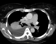
- Title:
- CT (chest), Pleural Effusion
- Description:
- Demonstrates associated pleural effusion and pleural thickening at the posterior aspect of the R lung (compare to healthy left lung). Note the part of the mass anterior to the effusion (hyperdense relative to the effusion).
- Keyword:
- Tomography, Ultrasonic, Echotomography, Echotomography, Computer, Computer Echotomography, Sonography, Medical, Ultrasonic Tomography, Echography, Pleura, Diagnosis, Ultrasonic Diagnosis, Ultrasonic Imaging, Ultrasound Imaging, Effusion, Diagnosis, Ultrasonic
- Subject:
- Diagnostic Techniques and Procedures, Diagnostic Imaging, Pleural Effusion, Ultrasonography, Multimodal Imaging
- Creator:
- Rush Medical College
- Publisher:
- Rush Medical College
- Language:
- English
- Rights:
- http://www.i-human.com/service-agreement-print
- Resource Type:
- Medical Imaging
- Identifier:
- 3225
- Title:
- CT (chest), Pleural Effusion
- Description:
- Demonstrates associated pleural effusion and pleural thickening at the posterior aspect of the R lung (compare to healthy left lung). Note the part of the mass anterior to the effusion (hyperdense relative to the effusion).
- Keyword:
- Effusion, Ultrasonic Diagnosis, Ultrasound Imaging, Diagnosis, Echotomography, Tomography, Ultrasonic, Echography, Sonography, Medical, Echotomography, Computer, Diagnosis, Ultrasonic, Pleura, Ultrasonic Tomography, Computer Echotomography, Ultrasonic Imaging
- Subject:
- Multimodal Imaging, Diagnostic Imaging, Diagnostic Techniques and Procedures, Pleural Effusion, Ultrasonography
- Creator:
- Rush Medical College
- Publisher:
- Rush Medical College
- Language:
- English
- Rights:
- http://www.i-human.com/service-agreement-print
- Resource Type:
- Medical Imaging
- Identifier:
- 3225
- Title:
- X-ray (chest), AP Lat - Pleural Effusion
- Description:
- There is a large opacity in the right lung base consistent with a large right pleural effusion. This was not present at the time of the comparison radiograph.
The radiographic appearance of the chest is otherwise unremarkable. The heart is normal in size. The aerated portions of both lungs appear normal. The left hemidiaphragm is well-defined, as is the left costophrenic sulcus.
IMPRESSION: New, large, right pleural effusion.
- Keyword:
- Ultrasonic Tomography, Sonography, Medical, Diagnosis, Ultrasonic, Diagnosis, Echography, Echotomography, Computer, Computer Echotomography, Tomography, Ultrasonic, Pleura, Effusion, Ultrasonic Diagnosis, Echotomography, Ultrasonic Imaging, Ultrasound Imaging
- Subject:
- Ultrasonography, Diagnostic Techniques and Procedures, Pleural Effusion, Diagnostic Imaging, Multimodal Imaging
- Creator:
- Rush Medical College
- Publisher:
- Rush Medical College
- Language:
- English
- Rights:
- http://www.i-human.com/service-agreement-print
- Resource Type:
- Medical Imaging
- Identifier:
- 3223
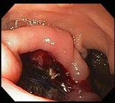
- Title:
- Endoscopy - Aortoenteric Fistula
- Description:
- Aortoenteric fistula
- Keyword:
- endoscopy, Minimally Invasive Surgical Procedures, Diagnosis
- Subject:
- Surgical Procedures, Operative, Aorta, Endoscopy, Biopsy, Diagnostic Techniques and Procedures, Diagnostic Techniques, Surgical
- Creator:
- Mike Sullivan MD Tufts Medical Center/ Tufts University School of Medicine
- Contributor:
- Tufts Medical Center
- Publisher:
- Mike Sullivan MD Tufts Medical Center/ Tufts University School of Medicine
- Language:
- English
- Rights:
- http://www.i-human.com/service-agreement-print
- Resource Type:
- Photo
- Identifier:
- 3215
