Search
« Previous |
181 - 190 of 960
|
Next »
Search Results
- Title:
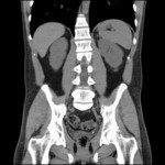
- CT (abdomen), Small Stone at the Left Ureteropelvic Junction
- Description:
- CT scan of abdomen without contrast demonstrating a small stone at the left ureteropelvic junction
- Keyword:
- Tomography, Ultrasonic, Diagnosis, Ultrasonic, Echography, kidney stone, Echotomography, Computer, Diagnosis, kidney stones, Ultrasonic Imaging, Ultrasound Imaging, Ultrasonic Tomography, Echotomography, Computer Echotomography, Ultrasonic Diagnosis, Sonography, Medical
- Subject:
- Diagnostic Techniques and Procedures, Ultrasonography, Male Urogenital Diseases, Diagnostic Imaging, Urologic Diseases, Multimodal Imaging, Kidney Diseases, Nephrolithiasis
- Creator:
- West Virginia University Department of Radiology
- Publisher:
- West Virginia University Department of Radiology
- Language:
- English
- Rights:
- http://www.i-human.com/service-agreement-print
- Resource Type:
- Medical Imaging
- Identifier:
- 3095
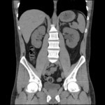
- Title:
- CT (abdomen), Mild Dilation of the Left Renal Collecting System
- Description:
- CT scan of abdomen with contrast demonstrating mild dilation of the left renal collecting system
- Keyword:
- Ultrasound Imaging, Ultrasonic Imaging, Ultrasonic Diagnosis, Sonography, Medical, kidney stone, Echotomography, Diagnosis, kidney stones, Computer Echotomography, Diagnosis, Ultrasonic, Echotomography, Computer, Tomography, Ultrasonic, Echography, Ultrasonic Tomography
- Subject:
- Kidney Diseases, Nephrolithiasis, Urologic Diseases, Multimodal Imaging, Diagnostic Techniques and Procedures, Ultrasonography, Diagnostic Imaging, Male Urogenital Diseases
- Creator:
- West Virginia University Department of Radiology
- Publisher:
- West Virginia University Department of Radiology
- Language:
- English
- Rights:
- http://www.i-human.com/service-agreement-print
- Identifier:
- 3094
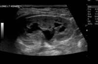
- Title:
- Ultrasound (abdomen), Hydronephrosis, Left Kidney
- Description:
- Single-image ultrasound demonstrating hydronephrosis of the left kidney
- Keyword:
- anatomy, Diagnosis, morphology, kidney, kidneys, Anatomy & histology, histology
- Subject:
- Nephrons, Multimodal Imaging, Kidney, Diagnostic Imaging, Diagnostic Techniques and Procedures, Ultrasonography, Urogenital System, Urinary Tract
- Creator:
- West Virginia University Department of Radiology
- Publisher:
- West Virginia University Department of Radiology
- Language:
- English
- Rights:
- http://www.i-human.com/service-agreement-print
- Resource Type:
- Medical Imaging
- Identifier:
- 3093
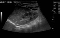
- Title:
- Ultrasound (abdomen), Normal, Right Kidney
- Description:
- Single-image ultrasound demonstrating a normal right kidney
- Keyword:
- histology, anatomy, Anatomy & histology, kidney, Diagnosis, kidneys, morphology
- Subject:
- Urinary Tract, Urogenital System, Multimodal Imaging, Diagnostic Techniques and Procedures, Kidney, Diagnostic Imaging, Nephrons, Ultrasonography
- Creator:
- West Virginia University Department of Radiology
- Publisher:
- West Virginia University Department of Radiology
- Language:
- English
- Rights:
- http://www.i-human.com/service-agreement-print
- Resource Type:
- Medical Imaging
- Identifier:
- 3092
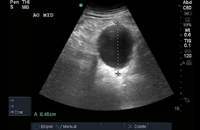
- Title:
- Ultrasound (abdomen), Abdominal Aortic Aneurysm
- Description:
- Single-image abdominal ultrasound demonstrating an 8.45-cm abdominal aortic aneurysm
- Keyword:
- Diagnosis, Computer Echotomography, Ultrasonic Tomography, Ultrasonic Imaging, Echotomography, Abdominal Aortic Aneurysm, Aneurysm, Echotomography, Computer, Sonography, Medical, Tomography, Ultrasonic, Ultrasonic Diagnosis, Ultrasound Imaging, Vascular Diseases, AAA, Diagnosis, Ultrasonic, Echography, Abdominal Aortic
- Subject:
- Multimodal Imaging, Cardiovascular Diseases, Ultrasonography, Aneurysm, Diagnostic Techniques and Procedures, Aortic Aneurysm, Diagnostic Imaging
- Creator:
- West Virginia University Department of Radiology
- Publisher:
- West Virginia University Department of Radiology
- Language:
- English
- Rights:
- http://www.i-human.com/service-agreement-print
- Resource Type:
- Medical Imaging
- Identifier:
- 3096
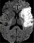
- Title:
- MRI (brain), Diffusion-Weighted Imaging, Left MCA Infarction
- Description:
- MRI with diffuse-weighted imaging demonstrating a left MCA infarction
- Keyword:
- CT X Ray, Diagnosis, Tomography, X Ray Computed, Computerized Tomography, X-Ray, Tomography, X-Ray Computerized, Computerized Tomography, X Ray, X Ray Tomography, Computed, Cine-CT, Tomography, Transmission Computed, CT Scan, X-Ray, Electron Beam Computed Tomography, X-Ray Computerized Axial Tomography, X-Ray Computer Assisted Tomography, Tomography, Xray Computed, Computed X Ray Tomography, Tomodensitometry, Computed Tomography, X-Ray, Electron Beam Tomography, CAT Scan, X Ray, CAT Scan, X-Ray, Acute Infarction - Right MCA, Tomography, X-Ray Computerized Axial, X-Ray Tomography, Computed, infarction, brain, MCA, CT, Cerebral,, Tomography, X-Ray Computer Assisted, X Ray Computerized Tomography
- Subject:
- Pathologic Processes, Diagnostic Imaging, Ischemia, Multimodal Imaging, Diagnostic Techniques and Procedures, Infarction, Pathological Conditions, Signs and Symptoms, Tomography, X-Ray Computed
- Creator:
- Radiology Assistant (reproducible, open source images for educational purposes with attribution) Brain Ischemia: Imaging in Acute Stroke Majda Thurnher, MD Medical University of Vienna
- Publisher:
- Radiology Assistant (reproducible, open source images for educational purposes with attribution) Brain Ischemia: Imaging in Acute Stroke Majda Thurnher, MD Medical University of Vienna
- Language:
- English
- Rights:
- http://www.i-human.com/service-agreement-print
- Resource Type:
- Medical Imaging
- Identifier:
- 3088
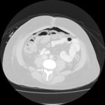
- Title:
- CT (abdomen), Free Intraperitoneal Air
- Description:
- Single-image CT of abdomen demonstrating free intraperitoneal air
- Keyword:
- X Ray Tomography, Computed, X-Ray Tomography, Computed, Tomography, X-Ray Computerized Axial, Tomography, X Ray Computed, Renal infarction, CT Scan, X-Ray, CAT Scan, X Ray, Computed Tomography, X-Ray, Infarction, X Ray Computerized Tomography, Tomography, X-Ray Computer Assisted, Electron Beam Computed Tomography, Tomography, Xray Computed, Computed X Ray Tomography, X-Ray Computer Assisted Tomography, Computerized Tomography, X Ray, Diagnosis, Tomodensitometry, Computerized Tomography, X-Ray, Kidney, CAT Scan, X-Ray, Electron Beam Tomography, Tomography, Transmission Computed, Cine-CT, X-Ray Computerized Axial Tomography, CT X Ray, Tomography, X-Ray Computerized
- Subject:
- Pathological Conditions, Signs and Symptoms, Infarction, Ischemia, Pathologic Processes, Multimodal Imaging, Diagnostic Imaging, Tomography, X-Ray Computed, Diagnostic Techniques and Procedures
- Creator:
- West Virginia University Department of Radiology
- Publisher:
- West Virginia University Department of Radiology
- Language:
- English
- Rights:
- http://www.i-human.com/service-agreement-print
- Resource Type:
- Medical Imaging
- Identifier:
- 3076
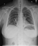
- Title:
- X-ray (chest), Pneumoperitoneum
- Description:
- Single-view CXR demonstrating free abdominal air
- Keyword:
- Diagnosis, Radiology, Diagnostic X-Ray, COPD, Chronic Obstructive Pulmonary Disease, Roentgenography, X-Ray Radiology, Diagnostic, Chronic Obstructive Lung Disease, Chronic Obstructive Airway Disease, Diagnostic X-Ray Radiology, COAD, Diagnostic X-Ray, X-Ray, Diagnostic, Chronic Airflow Obstruction
- Subject:
- Diagnostic Imaging, Lung Diseases, Obstructive, Lung Diseases, Pulmonary Disease, Chronic Obstructive, Respiratory Tract Diseases, Multimodal Imaging, Radiography, Diagnostic Techniques and Procedures
- Creator:
- West Virginia University Department of Radiology
- Publisher:
- West Virginia University Department of Radiology
- Language:
- English
- Rights:
- http://www.i-human.com/service-agreement-print
- Resource Type:
- Medical Imaging
- Identifier:
- 3075
- Title:
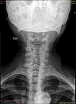
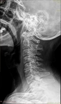
- X-ray (neck), Cervical Arthritis
- Description:
- Decreased bone density
Vertebral height and shape within normal parameters
Decreased space between C5-C6 and C6-C7. This change is most notorious in the first one, associated with surface sclerosis and incipient osteophytes.
Mild retrolisthesis on C5 which doesnt change significantly on dynamic projections. Sclerosis of the unciform apophysis C5 and C6. Other signs of sclerosis in C4-C5, C5-C6, and C6-C7.
Dg: Osteopenia; osteochondrosis C5-C6, C6-C7 associated with uncoarthrosis and spondyloarthrosis
- Keyword:
- joint, arthritis, Inflammation
- Subject:
- Musculoskeletal Diseases, Joint Diseases, Arthritis
- Creator:
- Jaime Drewes MD
- Publisher:
- Jaime Drewes MD
- Language:
- English
- Rights:
- http://www.i-human.com/service-agreement-print
- Resource Type:
- Medical Imaging
- Identifier:
- 3074
- Title:
- X-ray (neck), Cervical Arthritis
- Description:
- Decreased bone density
Vertebral height and shape within normal parameters
Decreased space between C5-C6 and C6-C7. This change is most notorious in the first one, associated with surface sclerosis and incipient osteophytes.
Mild retrolisthesis on C5 which doesnt change significantly on dynamic projections. Sclerosis of the unciform apophysis C5 and C6. Other signs of sclerosis in C4-C5, C5-C6, and C6-C7.
Dg: Osteopenia; osteochondrosis C5-C6, C6-C7 associated with uncoarthrosis and spondyloarthrosis
- Keyword:
- joint, Inflammation, arthritis
- Subject:
- Musculoskeletal Diseases, Joint Diseases, Arthritis
- Creator:
- Jaime Drewes MD
- Publisher:
- Jaime Drewes MD
- Language:
- English
- Rights:
- http://www.i-human.com/service-agreement-print
- Resource Type:
- Medical Imaging
- Identifier:
- 3074
