Search
« Previous |
101 - 110 of 120
|
Next »
Search Results
Select an image to start the slideshow
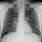
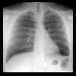
X-ray (chest), PA, Normal Inspiration and Expiration
1 of 10
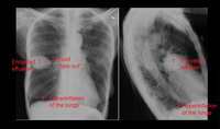
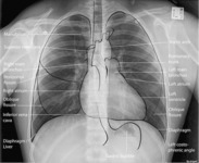
X-ray (chest), PA and Lateral, Encysted Effusion with Answers, Adult Male
2 of 10
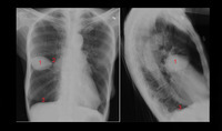
X-ray (chest), PA and Lateral, Encysted Effusion with Numbers, Adult Male
3 of 10
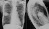
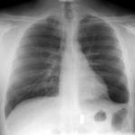
X-ray (chest), PA and Lateral, Encysted Effusion, Adult Male
4 of 10
X-ray (chest), AP, Adult Male, Thoracic Bullet, Annotated
5 of 10
X-ray (chest), PA, With Annotations, Adult Male, Normal
6 of 10
X-ray (chest), LAT, Adult Male, Thoracic Bullet
7 of 10
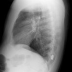
X-ray (chest), LAT, Adult Male, Thoracic Bullet
8 of 10
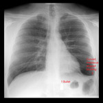
X-ray (chest), AP, Adult Male, Thoracic Bullet, Annotated Answers
9 of 10
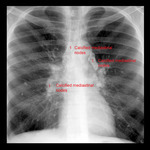
X-ray (chest), PA, Calcified Mediastinal Nodes, Adult Male
10 of 10
