Search
« Previous |
41 - 50 of 82
|
Next »
Search Results
- Title:
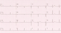
- 12-Lead ECG: Sinus bradycardia
- Description:
- HR: 51
QRS: 88 ms
Axis: P 79; QRS 68; T 67
- Keyword:
- Bradycardias, Bradyarrhythmias, Electrocardiograph, Diagnosis, EKG, ECG, Beta Blocker Overdose, Electrocardiogram, Heart
- Subject:
- Heart Diseases, Arrhythmias, Cardiac, Electrocardiography, Cardiovascular Diseases, Diagnostic Techniques, Cardiovascular, Bradycardia, Diagnostic Techniques and Procedures
- Publisher:
- i-Human Patients, Inc.
- Language:
- English
- Copyright Holder:
- i-Human Patients, Inc.
- Rights:
- http://www.i-human.com/service-agreement-print
- Resource Type:
- Chart/Diagram
- Identifier:
- 1820
- Title:
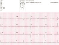
- 12-Lead ECG: Sinus bradycardia
- Description:
- HR: 51
QRS: 88 ms
Axis: P 79; QRS 68; T 67
- Keyword:
- Heart, Bradycardias, Bradyarrhythmias, Electrocardiograph, ECG, Diagnosis, EKG, Beta Blocker Overdose, Electrocardiogram
- Subject:
- Diagnostic Techniques and Procedures, Bradycardia, Diagnostic Techniques, Cardiovascular, Arrhythmias, Cardiac, Heart Diseases, Electrocardiography, Cardiovascular Diseases
- Publisher:
- i-Human Patients, Inc.
- Language:
- English
- Copyright Holder:
- i-Human Patients, Inc.
- Rights:
- http://www.i-human.com/service-agreement-print
- Resource Type:
- Chart/Diagram
- Identifier:
- 1820
- Title:
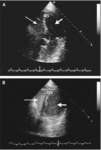
- Transthoracic Echocardiogram (TTE), Atrial Septal Defect (ASD)
- Description:
- The image in Panel A shows minimal right-to-left shunting through the patent foramen ovale (arrow). It was obtained with the patient in the supine position.
The image in Panel B shows increased right-to-left shunting (arrow). It was obtained with the patient in the sitting position. LA denotes left atrium, RA right atrium, and Ao aorta.
- Keyword:
- Echocardiography, M-Mode, Transthoracic Echocardiography, Echocardiography, 2-D, Cross-Sectional Echocardiography, 2D Echocardiography, Atrial Septal Defects, Echocardiography, Cross-Sectional, Heart, Echocardiography, Two-Dimensional, Persistent Ostium Primum, Diagnosis, Echocardiography, 2D, Echocardiography, Contrast, Atrial Septal Defect, Echocardiography, Transthoracic, Ostium Secundum Atrial Septal Defect, 2-D Echocardiography, M-Mode Echocardiography, Two-Dimensional Echocardiography, Contrast Echocardiography
- Subject:
- Cardiac Imaging Techniques, Multimodal Imaging, Echocardiography, Heart Defects, Congenital, Cardiovascular Diseases, Diagnostic Imaging, Cardiovascular Abnormalities, Diagnostic Techniques and Procedures, Heart Septal Defects
- Creator:
- Paul Kent, M.D. Rush Medical College
- Publisher:
- i-Human Patients, Inc.
- Language:
- English
- Copyright Holder:
- Paul Kent, M.D.
- Rights:
- http://www.i-human.com/service-agreement-print
- Resource Type:
- Medical Imaging
- Identifier:
- 1802
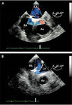
- Title:
- Transthoracic Echocardiogram (TTE), Atrial Septal Defect (ASD)
- Description:
- Note the passage of bubbles from the RA to the LA and flow (blue towards the transducer, red away) on the Doppler echo.
- Keyword:
- 2D Echocardiography, Atrial Septal Defect, Persistent Ostium Primum, Echocardiography, 2-D, Echocardiography, Cross-Sectional, Ostium Secundum Atrial Septal Defect, M-Mode Echocardiography, Two-Dimensional Echocardiography, 2-D Echocardiography, Echocardiography, M-Mode, Echocardiography, Two-Dimensional, Echocardiography, 2D, Transthoracic Echocardiography, Cross-Sectional Echocardiography, Echocardiography, Transthoracic, Heart, Diagnosis, Atrial Septal Defects, Contrast Echocardiography, Echocardiography, Contrast
- Subject:
- Echocardiography, Diagnostic Techniques and Procedures, Diagnostic Imaging, Cardiac Imaging Techniques, Multimodal Imaging, Heart Septal Defects, Cardiovascular Abnormalities, Heart Defects, Congenital, Cardiovascular Diseases
- Creator:
- Paul Kent, M.D.
- Publisher:
- i-Human Patients, Inc.
- Language:
- English
- Copyright Holder:
- Paul Kent, M.D.
- Rights:
- http://www.i-human.com/service-agreement-print
- Resource Type:
- Medical Imaging
- Identifier:
- 1798
- Title:
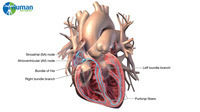
- Cardiac Conduction; Labels
- Description:
- Illustration - Cardiac conduction; label and matching still
- Keyword:
- Right Bundle Branch, Bundle of His, Heart Block, AV node, Purkinje Fibers, Left Bundle Branch, Heart, Arrhythmias, SA node
- Subject:
- Bundle of His, Purkinje Fibers, Cardiovascular System, Heart Conduction System, Sinoatrial Node, Atrioventricular Node, Bundle-Branch Block
- Creator:
- Laura Garrison, MS
- Publisher:
- i-Human Patients; Inc.
- Language:
- English
- Copyright Holder:
- i-Human Patients, Inc.
- Rights:
- http://www.i-human.com/service-agreement-print
- Resource Type:
- Illustration
- Title:
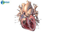
- Cardiac Conduction; Matching Exercise
- Description:
- Animation - Cardiac conduction; label and matching video and stills
- Keyword:
- AV node, Heart, Bundle of His, Left Bundle Branch, Arrhythmias, SA node, Heart Block, Right Bundle Branch, Purkinje Fibers
- Subject:
- Atrioventricular Node, Bundle-Branch Block, Heart Conduction System, Sinoatrial Node, Bundle of His, Cardiovascular System, Purkinje Fibers
- Creator:
- Laura Garrison
i-Human Patients; Inc.
- Language:
- English
- Copyright Holder:
- i-Human Patients, Inc.
- Rights:
- http://www.i-human.com/service-agreement-print
- Resource Type:
- Video
- Title:
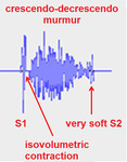
- Murmur, Aortic stenosis
- Description:
- Murmur - Aortic stenosis. Thinning of the aortic valve due to age, inborn error (biscuspid aortic valve), or metastatic calcification of the valve
- Keyword:
- Heart Ventricles, Heart, Valve Stenoses, Aortic, Stenosis, Aortic, Valve Stenosis, Aortic, Stenoses, Aortic Valve, Heart Diseases, Aortic Valve Stenoses, Stenoses, Aortic, Stenosis, Aortic Valve, Aortic Stenosis
- Subject:
- Cardiovascular Diseases, Ventricular Outflow Obstruction, Heart Valve Diseases, Aorta, Aortic Valve Stenosis
- Creator:
- Avery Ellis, M.D., Ph.D.
Associate Professor Cardiology
Senior Associate Dean Medicine
University at Buffalo School of Medicine
- Publisher:
- University of Buffalo School of Medicine
- Language:
- English
- Copyright Holder:
- Albert Einstein College of Medicine
- Rights:
- http://www.i-human.com/service-agreement-print
- Resource Type:
- Chart/Diagram
- Identifier:
- 1743
- Title:
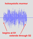
- Murmur, Mitral Regurgitation
- Description:
- Murmur - Mitral Regurgitation
- Keyword:
- Heart, Valve Regurgitation, Mitral, Mitral Incompetence, Regurgitation, Mitral, Valve Insufficiency, Mitral, Mitral Valve Incompetence, Incompetence, Mitral Valve, Mitral Valve Regurgitation, Regurgitation, Mitral Valve, Heart Ventricles, Insufficiency, Mitral Valve, Incompetence, Mitral, Valve Incompetence, Mitral, Mitral Insufficiency, Heart Diseases, Insufficiency, Mitral, Mitral Regurgitation
- Subject:
- Mitral Valve Insufficiency, Heart Valve Diseases, Ventricular Outflow Obstruction, Aorta, Cardiovascular Diseases
- Creator:
- Avery Ellis, M.D., Ph.D.
Associate Professor Cardiology
Senior Associate Dean Medicine University at Buffalo School of Medicine
- Publisher:
- University of Buffalo School of Medicine
- Language:
- English
- Copyright Holder:
- Albert Einstein College of Medicine
- Rights:
- http://www.i-human.com/service-agreement-print
- Resource Type:
- Chart/Diagram
- Identifier:
- 1744
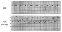
- Title:
- 3 Lead ECG with pulmonary capillary wedge pressure (PCW)
- Description:
- 3 Lead ECG with elevated pulmonary capillary wedge pressure (PCW)
- Keyword:
- Electrocardiogram, Wedge Pressures, Pulmonary, Wedge Pressure, Pulmonary, Pulmonary Wedge Pressure, Pulmonary Capillary Wedge Pressure, ECG, Diagnosis, Heart, Wedge Pressures, Pulmonary Venous Wedge Pressure, Wedge Pressure, Electrocardiograph, Pulmonary Wedge Pressures, Pressure, Wedge, Pressures, Wedge, EKG, Pressures, Pulmonary Wedge, Pulmonary Artery Wedge Pressure, Pressure, Pulmonary Wedge
- Subject:
- Circulatory and Respiratory Physiological Phenomena, Electrocardiography, Diagnostic Techniques and Procedures, Diagnostic Techniques, Cardiovascular, Cardiovascular Physiological Phenomena, Blood Pressure, Hemodynamics
- Creator:
- Avery Ellis, M.D., Ph.D.
University of Buffalo School of Medicine
- Publisher:
- University of Buffalo School of Medicine
- Language:
- English
- Copyright Holder:
- University at Buffalo School of Medicine
- Rights:
- http://www.i-human.com/service-agreement-print
- Resource Type:
- Chart/Diagram
- Identifier:
- 1707
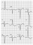
- Title:
- ECG: Left ventricular hypertrophy (LVH) with left ventricular dilation (LAD)
- Description:
- ECG left ventricular hypertrophy (LVH) with left ventricular dilation (LAD)
- Keyword:
- Left Ventricular Hypertrophy, Electrocardiograph, Hypertrophies, Left Ventricular, EKG, Electrocardiogram, Ventricular Hypertrophy, Left, Heart, Ventricular Hypertrophies, Left, Diagnosis, ECG, Left Ventricular Hypertrophies
- Subject:
- Pathological Conditions, Anatomical, Diagnostic Techniques, Cardiovascular, Diagnostic Techniques and Procedures, Cardiomegaly, Hypertrophy, Left Ventricular, Hypertrophy, Electrocardiography
- Creator:
- Avery Ellis, M.D., PhD, University of Buffalo School of Medicine
- Publisher:
- i-Human Patients, Inc.
- Language:
- English
- Copyright Holder:
- University of Buffalo School of Medicine
- Rights:
- http://www.i-human.com/service-agreement-print
- Resource Type:
- Chart/Diagram
- Identifier:
- 1709
