Search
« Previous |
1 - 10 of 18
|
Next »
Search Results
Select an image to start the slideshow

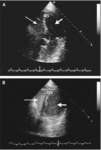
Transthoracic echocardiogram (TTE), Hypertrophic cardiomyopathy (HCM) (4CH)
1 of 10
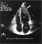
Transthoracic echocardiogram (TEE): Apical Hypertrophic cardiomyopathy 4CH
2 of 10
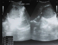
Ultrasound (abdomen), Liver Abscess
3 of 10
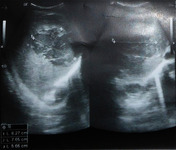
Ultrasound (abdomen), Liver Abscess
4 of 10
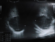
Ultrasound (abdomen), Liver Abscess
5 of 10
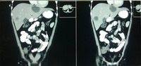
CT (abdomen), Liver Abscess
6 of 10
Transthoracic Echocardiogram (TTE), Atrial Septal Defect (ASD)
7 of 10
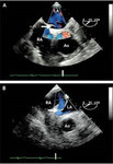
Transthoracic Echocardiogram (TTE), Atrial Septal Defect (ASD)
8 of 10
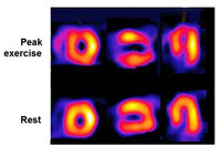
Nuclear Stress Test
9 of 10
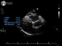
Transthoracic Echocardiogram (TTE), Left Ventricular Hypertrophy (LAX)
10 of 10
