Search
« Previous |
181 - 190 of 341
|
Next »
Search Results
- Title:
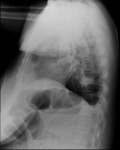
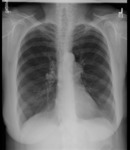
- X-ray (chest Lateral), Granulomatosis with Polyangiitis (Wegeners Granulomatosis)
- Description:
- Wegeners granulomatosis
Adult female
Granulomatosis with polyangiitis (GPA)
Multiple b/l nodular opacities >5 cm in diam right mid-lung field and apices bilaterally; normal cor size; no effusion
- Keyword:
- Radiography, X-Ray, Diagnostic, Radiology, Diagnostic X-Ray, Wegener Granulomatosis, Diagnosis, Diagnostic X-Ray Radiology, Wegener's Granulomatosis, Diagnostic X-Ray, X-Ray Radiology, Diagnostic, Granulomatosis, Wegener's, Roentgenography
- Subject:
- Granulomatosis with Polyangiitis, Multimodal Imaging, Autoimmune Diseases, Diagnostic Imaging, Immune System Diseases, Anti-Neutrophil Cytoplasmic Antibody-Associated Vasculitis, Diagnostic Techniques and Procedures
- Creator:
- i-Human Patients, Inc., Inc.
- Publisher:
- i-Human Patients, Inc., Inc.
- Language:
- English
- Copyright Holder:
- i-Human Patients, Inc.
- Rights:
- http://www.i-human.com/service-agreement-print
- Identifier:
- 1721
- Title:
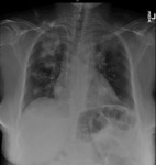
- X-ray (chest PA), Granulomatosis with Polyangiitis (Wegeners Granulomatosis)
- Description:
- Wegeners granulomatosis
Adult Female, CXR PA
Granulomatosis with polyangiitis (GPA)
Multiple b/l nodular opacities >5 cm in diam right mid-lung field and apices bilaterally; normal cor size; no effusion
- Keyword:
- X-Ray Radiology, Diagnostic, Roentgenography, Wegener's Granulomatosis, Radiology, Diagnostic X-Ray, Granulomatosis, Wegener's, Wegener Granulomatosis, X-Ray, Diagnostic, Radiography, Diagnosis, Diagnostic X-Ray, Diagnostic X-Ray Radiology
- Subject:
- Anti-Neutrophil Cytoplasmic Antibody-Associated Vasculitis, Diagnostic Techniques and Procedures, Autoimmune Diseases, Immune System Diseases, Diagnostic Imaging, Granulomatosis with Polyangiitis, Multimodal Imaging
- Creator:
- i-Human Patients, Inc., Inc.
- Publisher:
- i-Human Patients, Inc., Inc.
- Language:
- English
- Copyright Holder:
- i-Human Patients, Inc.
- Rights:
- http://www.i-human.com/service-agreement-print
- Identifier:
- 1721
- Title:
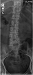
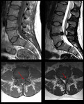
- X-ray (lumbar spine), Coronal and Sagital Views
- Description:
- Views of the lumbar spine demonstrate preservation of the vertebrae with normal alignment and no fractures. No boney lesions noted but there is the suggestion of diminished bone density.
- Keyword:
- Diagnosis, Radiology, Diagnostic X-Ray, Roentgenography, Radiography, Spine, Diagnostic X-Ray, Diagnostic X-Ray Radiology, Musculoskeletal Diseases, X-Ray Radiology, Diagnostic, X-Ray, Diagnostic
- Subject:
- Multimodal Imaging, Diagnostic Imaging, Spine, Bone and Bones, Skeleton, Diagnostic Techniques and Procedures
- Creator:
- Jah-Won Koo, MDRush Medical College
- Publisher:
- Rush Medical College
- Language:
- English
- Copyright Holder:
- Rush Medical College
- Rights:
- http://www.i-human.com/service-agreement-print
- Resource Type:
- Medical Imaging
- Identifier:
- 1719

- Title:
- X-ray (lumbar spine), Coronal and Sagital Views
- Description:
- Views of the lumbar spine demonstrate preservation of the vertebrae with normal alignment and no fractures. No boney lesions noted but there is the suggestion of diminished bone density.
- Keyword:
- X-Ray, Diagnostic, Diagnostic X-Ray Radiology, Radiology, Diagnostic X-Ray, Musculoskeletal Diseases, Diagnostic X-Ray, Radiography, Spine, X-Ray Radiology, Diagnostic, Roentgenography, Diagnosis
- Subject:
- Diagnostic Imaging, Bone and Bones, Spine, Multimodal Imaging, Skeleton, Diagnostic Techniques and Procedures
- Creator:
- Jah-Won Koo, MDRush Medical College
- Publisher:
- Rush Medical College
- Language:
- English
- Copyright Holder:
- Rush Medical College
- Rights:
- http://www.i-human.com/service-agreement-print
- Resource Type:
- Medical Imaging
- Identifier:
- 1719
- Title:
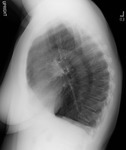
- X-ray (chest), PA and Lateral, Chronic Obstructive Pulmonary Disease (COPD), Adult Female
- Description:
- Posterior-anterior and Lateral views of the chest reveal clear lung fields, normal cardiac silhouette, but with hyperexpansion and flattened diaphragms consistent with obstructive airways disease such as emphysema or asthma. No infiltrate, mass, or pulmonary vascular congestion is seen.
- Keyword:
- X-Ray, Diagnostic, Radiology, Diagnostic X-Ray, Chronic Obstructive Airway Disease, Radiography, Diagnostic X-Ray Radiology, Diagnostic X-Ray, COPD, Roentgenography, Diagnosis, X-Ray Radiology, Diagnostic, Chronic Airflow Obstruction, Chronic Obstructive Lung Disease, Chronic Obstructive Pulmonary Disease, COAD
- Subject:
- Diagnostic Techniques and Procedures, Respiratory Tract Diseases, Lung Diseases, Lung Diseases, Obstructive, Multimodal Imaging, Pulmonary Disease, Chronic Obstructive, Diagnostic Imaging
- Creator:
- Jah-Won Koo, MDRush Medical College
- Publisher:
- Rush Medical College
- Language:
- English
- Copyright Holder:
- Rush Medical College
- Rights:
- http://www.i-human.com/service-agreement-print
- Resource Type:
- Medical Imaging
- Identifier:
- 1718
- Title:
- X-ray (chest), PA and Lateral, Chronic Obstructive Pulmonary Disease (COPD), Adult Female
- Description:
- Posterior-anterior and Lateral views of the chest reveal clear lung fields, normal cardiac silhouette, but with hyperexpansion and flattened diaphragms consistent with obstructive airways disease such as emphysema or asthma. No infiltrate, mass, or pulmonary vascular congestion is seen.
- Keyword:
- Roentgenography, Diagnosis, Chronic Obstructive Lung Disease, Diagnostic X-Ray Radiology, Diagnostic X-Ray, Chronic Obstructive Pulmonary Disease, Radiology, Diagnostic X-Ray, Radiography, COAD, Chronic Airflow Obstruction, X-Ray, Diagnostic, COPD, Chronic Obstructive Airway Disease, X-Ray Radiology, Diagnostic
- Subject:
- Respiratory Tract Diseases, Diagnostic Techniques and Procedures, Lung Diseases, Obstructive, Multimodal Imaging, Pulmonary Disease, Chronic Obstructive, Lung Diseases, Diagnostic Imaging
- Creator:
- Jah-Won Koo, MDRush Medical College
- Publisher:
- Rush Medical College
- Language:
- English
- Copyright Holder:
- Rush Medical College
- Rights:
- http://www.i-human.com/service-agreement-print
- Resource Type:
- Medical Imaging
- Identifier:
- 1718
- Title:
- MRI, Sagittal and Transverse - Disc herniation
- Description:
- L4-L5 left paracentral disc herniation causing stenosis of the left lateral recess and compression of the traversing left L5 nerve root
- Keyword:
- Proton Spin Tomography, fMRI, NMR Tomography, Anatomy, Cross-Sectional, MRI Scans, Musculoskeletal Diseases, Functional Magnetic Resonance Imaging, Tomography, MR, spine, Disk Prolapse, Diagnosis, Tomography, Proton Spin, MRI, Functional, NMR Imaging, Prolapsed Disc, Chemical Shift Imaging, Tomography, NMR, MR Tomography, Slipped Disk, Intervertebral Disk Displacement, Slipped Disc, Disc, Herniated, Herniated Disc, Imaging, Chemical Shift, Magnetization Transfer Contrast Imaging, Magnetic Resonance Spectroscopy, Magnetic Resonance Imaging, Functional, Herniated Disk, Disk, Herniated, Prolapsed Disk, Zeugmatography
- Subject:
- Intervertebral Disc Displacement, Bone Diseases, Multimodal Imaging, Diagnostic Imaging, Magnetic Resonance Imaging, Spinal Diseases, Diagnostic Techniques and Procedures
- Creator:
- Jah-Won KooRush Medical College
- Publisher:
- Rush Medical College
- Language:
- English
- Copyright Holder:
- Rush Medical College
- Rights:
- http://www.i-human.com/service-agreement-print
- Identifier:
- 1720
- Title:
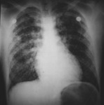
- X-ray (chest), AP, Pulmonary Infiltrate, Mitral Insufficiency / Regurgitation
- Description:
- The chest x-ray of an adult male shows a normal cardiac silhouette with a diffuse infiltrate throughout both lung fields suggesting pulmonary congestion secondary to mitral valve regurgitation.
- Keyword:
- pulmonary congestion, Diagnostic X-Ray Radiology, Roentgenography, Diagnosis, Radiography, Radiology, Diagnostic X-Ray, X-Ray, Diagnostic, pumonary infiltrate, X-Ray Radiology, Diagnostic, Diagnostic X-Ray
- Subject:
- Lung Diseases, Multimodal Imaging, Diagnostic Imaging, Respiratory Tract Diseases, Pulmonary Edema, Diagnostic Techniques and Procedures
- Creator:
- Avery Ellis, M.D., Ph.D.University of Buffalo School of Medicine
- Publisher:
- University of Buffalo School of Medicine
- Language:
- English
- Copyright Holder:
- University at Buffalo School of Medicine
- Rights:
- http://www.i-human.com/service-agreement-print
- Identifier:
- 1697

- Title:
- X-ray (chest), PA, Congestive Heart Failure (CHF), Adult Male
- Description:
- CXR PA - Adult male, congestive heart failure (CHF)
1) baseline (prior film)
2) on presentation
3) symptomatic
- Keyword:
- X-Ray, Diagnostic, Right-Sided Heart Failure, Heart Failure, Congestive, Heart Decompensation, X-Ray Radiology, Diagnostic, Diagnostic X-Ray, Congestive Heart Failure, Heart Failure, Left-Sided, Myocardial Failure, Diagnostic X-Ray Radiology, Heart Failure, Right-Sided, Roentgenography, Radiology, Diagnostic X-Ray, Diagnosis, Left-Sided Heart Failure, Cardiac Failure, Radiography, Heart Failure
- Subject:
- Multimodal Imaging, Diagnostic Imaging, Diagnostic Techniques and Procedures, Cardiovascular Diseases, Heart Diseases
- Creator:
- Avery Ellis, M.D., Ph.D. University of Buffalo School of Medicine
- Publisher:
- University of Buffalo School of Medicine
- Language:
- English
- Copyright Holder:
- University at Buffalo School of Medicine
- Rights:
- http://www.i-human.com/service-agreement-print
- Resource Type:
- Medical Imaging
- Identifier:
- 1712
- Title:
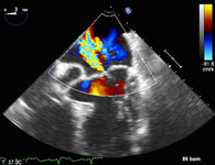
- Transesophageal Doppler Echocardiogram (TEE), Mitral Valve Prolapse, Mitral Regurgitation
- Description:
- Echocardiogram - Transthoracic echocardiogram (TTE)
MVP-MR LAX (2) and MVP-MR Color Doppler
- Keyword:
- Diagnosis, Echocardiography, Doppler Color, Transthoracic Echocardiography, Color Flow Echocardiography, Mitral Insufficiency, Echocardiography, Color Flow, Mitral Incompetence, M-Mode Echocardiography, Two-Dimensional Echocardiography, Color Doppler Echocardiography, Echocardiography, M-Mode, Prolapsed Mitral Valve, Doppler Echocardiography, Color, Mitral Valve Prolapse Syndrome, Mitral Click-Murmur Syndrome, Echocardiography, Contrast, Cross-Sectional Echocardiography, Echocardiography, 2-D, Echocardiography, Color Doppler, Mitral Valve Regurgitation, Mitral Valve Incompetence, Systolic Click-Murmur Syndrome, Echocardiography, Cross-Sectional, Echocardiography, Transthoracic, 2-D Echocardiography, Echocardiography, 2D, Floppy Mitral Valve, 2D Echocardiography, Mitral Regurgitation, Echocardiography, Two-Dimensional, Doppler Color Echocardiography, Contrast Echocardiography, Click-Murmur Syndrome, Color Echocardiography, Doppler
- Subject:
- Heart Valve Diseases, Multimodal Imaging, Diagnostic Techniques and Procedures, Ultrasonography, Mitral Valve Prolapse, Echocardiography, Doppler, Color, Cardiovascular Diseases, Heart Function Tests, Echocardiography, Transesophageal, Mitral Valve Insufficiency, Diagnostic Imaging, Heart Diseases, Echocardiography
- Creator:
- Avery Ellis, M.D., Ph.D. University of Buffalo School of Medicine
- Publisher:
- i-Human Patients, Inc.
- Language:
- English
- Copyright Holder:
- Avery Ellis, MD, PhD
- Rights:
- http://www.i-human.com/service-agreement-print
- Resource Type:
- Medical Imaging
- Identifier:
- 1699
