Search
« Previous |
1 - 10 of 14
|
Next »
Search Results
Select an image to start the slideshow
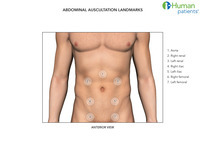
Auscultation landmarks, abdomen
1 of 10
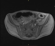
MRI: Ulcerative Colitis
2 of 10
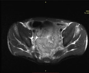
MRI: Ulcerative Colitis
3 of 10
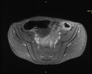
MRI: Ulcerative Colitis
4 of 10

Endoscopy, Mallory-Weiss tear
5 of 10
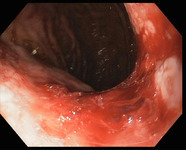
Endoscopy, Mallory-Weiss tear
6 of 10
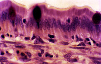
Small Intestine, Simple Columnar Epithelium, Cat
7 of 10
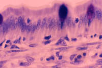
Small Intestine, Simple Columnar Epithelium, Cat
8 of 10

Small Intestine, Simple Columnar Epithelium, Cat
9 of 10
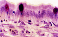
Small Intestine, Simple Columnar Epithelium, Cat
10 of 10
