Search

You searched for: File Format png (Portable Network Graphics) Remove constraint File Format: png (Portable Network Graphics) Subject Lung Remove constraint Subject: Lung

Search Results

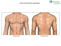
  1. Auscultation landmarks, lungs

    show more
    Description: Lung auscultation landmarks
    Keywords: Wheezes, Respiratory Sounds, Crackles, Stridor, Rales, Respiratory System, Auscultation, Ronchi
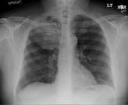
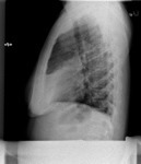
  2. X-ray (chest), AP and LAT: Lung Mass

    show more
    Description: Main Finding: This image shows a well define mass in the right upper lobe. The lateral view demonstrates that the mass is within the lung parenchyma in the right upper lobe. FINDINGS: A large right upper lobe mass is identified, corresponding to the known mass in this location. Right apical bulla are also seen. The trachea is not deviated. Otherwise there is no focal consolidation, pleural effusion, or pneumothorax. The cardiomediastinal silhouette and pulmonary vasculature are normal. Left basilar subsegmental atelectasis is noted. The regional skeleton is unremarkable. On the lateral view, the vertebral body heights appear preserved.
    Keywords: respiratory system, lung, Mice, morphology, histology, Anatomy & histology, lungs, anatomy
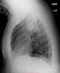
  3. X-ray (chest), AP and Lateral, Lung Mass

    show more
    Description: Main Finding: This image shows a well define mass in the right upper lobe. The lateral view demonstrates that the mass is within the lung parenchyma in the right upper lobe. FINDINGS: A large right upper lobe mass is identified, corresponding to the known mass in this location. Right apical bulla are also seen. The trachea is not deviated. Otherwise there is no focal consolidation, pleural effusion, or pneumothorax. The cardiomediastinal silhouette and pulmonary vasculature are normal. Left basilar subsegmental atelectasis is noted. The regional skeleton is unremarkable. On the lateral view, the vertebral body heights appear preserved.
    Keywords: Mice, anatomy, lung, Anatomy & histology, histology, respiratory system, morphology, lungs
  4. X-Ray (chest), AP and LAT, Adult Male, Normal

    show more
    Description: Test within normal limits
    Keywords: X-Ray Radiology, Diagnostic, X-Ray, Diagnostic, Radiology, Diagnostic X-Ray, Diagnostic X-Ray Radiology, Diagnosis, Diagnostic X-Ray
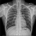
  5. X-ray (chest), AP and LAT, Adult Male, Normal

    show more
    Description: Test within normal limits
    Keywords: Diagnostic X-Ray Radiology, Radiology, Diagnostic X-Ray, Diagnosis, Diagnostic X-Ray, X-Ray Radiology, Diagnostic, X-Ray, Diagnostic
  6. X-ray (chest), Lungs, Atypical Pneumonia

    show more
    Description: Single radiographic image demonstrating an atypical pneumonia
    Keywords: Roentgenography, Radiography, X-Ray, Diagnostic, Diagnostic X-Ray Radiology, Radiology, Diagnostic X-Ray, X-Ray Radiology, Diagnostic, Diagnostic X-Ray
  7. X-ray (chest), Lateral, Right Middle Lobe Pneumonia

    show more
    Description: Single radiographic image demonstrating an atypical pneumonia Adult female
    Keywords: Radiography, X-Ray, Diagnostic, Roentgenography, Radiology, Diagnostic X-Ray, Diagnostic X-Ray, Diagnostic X-Ray Radiology, X-Ray Radiology, Diagnostic
  8. X-ray (chest), PA, Right Middle Lobe Pneumonia

    show more
    Description: Single radiographic image demonstrating an atypical pneumonia Adult female
    Keywords: Radiography, X-Ray, Diagnostic, Roentgenography, Diagnostic X-Ray Radiology, Diagnostic X-Ray, X-Ray Radiology, Diagnostic, Radiology, Diagnostic X-Ray
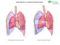
  9. Pneumothorax (Spontaneous Vs. Tension)

    show more
    Description: Spontaneous pneumothorax vs. tension pneumothorax
    Keywords: Respiratory Tract Diseases, Spontaneous Pneumothorax, Wounds and Injuries, Lung, Respiration Disorders, Tension Pneumothorax
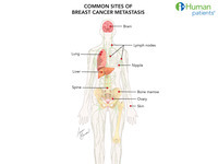
  10. Breast Cancer Metastases

    show more
    Description: Illustration of breast cancer metastases
    Keywords: Neoplasm Metastasis, Brain, Lung, Skin, Bone Marrow, Lymph Nodes, Breast Neoplasms, Ovary, Spine, Liver