Search
« Previous |
161 - 170 of 1,313
|
Next »
Search Results
- Title:
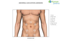
- Auscultation landmarks, abdomen
- Description:
- Abdominal auscultation landmarks
- Keyword:
- Digestive System, Intestine, Large, Physical Examinations, Examinations, Physical, Auscultation, Intestinal Obstruction, Obstruction, Examination, Physical, Physical Examinations and Diagnoses
- Subject:
- Colon, Gastroenteritis, Colitis, Abdominal Pain, Colonic Diseases, Intestines
- Creator:
- Kristina DeRyke, i-Human Patients, Inc.
- Publisher:
- i-Human Patients, Inc.
- Language:
- English
- Copyright Holder:
- i-Human Patients, Inc.
- Rights:
- http://www.i-human.com/service-agreement-print
- Resource Type:
- Illustration
- Identifier:
- 3579
- Title:
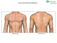
- Auscultation landmarks, lungs
- Description:
- Lung auscultation landmarks
- Keyword:
- Wheezes, Respiratory Sounds, Crackles, Stridor, Rales, Respiratory System, Auscultation, Ronchi
- Subject:
- Lung, Respiratory Sounds, Respiratory System
- Creator:
- Kristina DeRyke, i-Human Patients, Inc.
- Publisher:
- i-Human Patients, Inc.
- Language:
- English
- Copyright Holder:
- i-Human Patients, Inc.
- Rights:
- http://www.i-human.com/service-agreement-print
- Resource Type:
- Illustration
- Identifier:
- 3577
- Title:
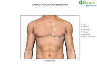
- Auscultation landmarks, heart
- Description:
- Cardiac auscultation landmarks
- Keyword:
- heart rate, beats per minute, cardiac sound, heart sound, cardiac auscultation sound, simulation, cardiac sounds, cardiac auscultation sounds, Heart, heart sounds
- Subject:
- Cardiovascular System, Heart Sounds
- Creator:
- Kristina DeRyke, i-Human Patients, Inc.
- Publisher:
- i-Human Patients, Inc.
- Language:
- English
- Rights:
- http://www.i-human.com/service-agreement-print
- Resource Type:
- Illustration
- Identifier:
- 3578
- Title:
- TEST - Shoulder_MusclesPosterior.png
- Description:
- Samples for testing purpose
- Subject:
- Phacoemulsification, Cataract Extraction, Refractive Surgical Procedures, Ophthalmologic Surgical Procedures, Surgical Procedures, Operative
- Creator:
- nilima@i-human.com
- Rights:
- http://creativecommons.org/licenses/by/3.0/us/
- Resource Type:
- Photograph

- Title:
- TEST - crosshatch.png
- Description:
- Samples for testing purpose
- Subject:
- Phacoemulsification, Cataract Extraction, Refractive Surgical Procedures, Ophthalmologic Surgical Procedures, Surgical Procedures, Operative
- Creator:
- nilima@i-human.com
- Rights:
- http://creativecommons.org/licenses/by/3.0/us/
- Resource Type:
- Photograph

- Title:
- TEST - crosshatch2.png
- Description:
- Samples for testing purpose
- Subject:
- Phacoemulsification, Cataract Extraction, Refractive Surgical Procedures, Ophthalmologic Surgical Procedures, Surgical Procedures, Operative
- Creator:
- nilima@i-human.com
- Rights:
- http://creativecommons.org/licenses/by/3.0/us/
- Resource Type:
- Photograph
- Title:
- TEST-cheetah_running.png
- Description:
- test upload
- Subject:
- Phacoemulsification, Cataract Extraction, Refractive Surgical Procedures, Ophthalmologic Surgical Procedures, Surgical Procedures, Operative
- Creator:
- nilima@i-human.com
- Rights:
- http://creativecommons.org/licenses/by/3.0/us/

- Title:
- TEST-Light_Pink_Rose.png
- Description:
- test upload
- Subject:
- Phacoemulsification, Cataract Extraction, Refractive Surgical Procedures, Ophthalmologic Surgical Procedures, Surgical Procedures, Operative
- Creator:
- nilima@i-human.com
- Rights:
- http://creativecommons.org/licenses/by/3.0/us/
- Title:
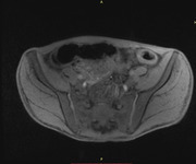
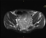
- MRI: Ulcerative Colitis
- Description:
- Main Feature: Large bowel wall thickening.
Description: There is diffuse circumferential wall thickening, hyperintense T2 signal, and avid mucosal enhancement throughout the entire colon and rectum. Increased mesenteric fat stranding surrounding the bowel with prominence of the mesenteric vessels. The terminal ileum is grossly unremarkable. These findings are suggestive of ulcerative colitis.
Series 30, image 5096-axial/t1, precontrast. This picture is to be used together with the series 39, image 5096. The point of focus here is the descending colon located at the 1-2 oclock position. Notice that the bowel wall is circumferentially thickened on both the pre and post contrast image. On series 39, you see that same portion of the colon wall diffusely enhance. Also, in series 39 note the engorged vessels that feed the colon.
Series 39, image 5096-axial/t1, postcontrast.
Series 24, image 29- t2 axial. At the level of the sigmoid colon. The purpose of this image is to show diffuse circumferential wall thickening in one continuous segment, hyperintense t2 signal, and mucosal enhancement.
- Keyword:
- hyperplasia, hyperplastic cells, Colitis, Ulcerative, cancer polyp
- Subject:
- Intestine, Large, Polyps, Intestines, Digestive System Diseases, Pathological Conditions, Anatomical, Gastroenteritis, Colon, Gastrointestinal Diseases, Intestinal Polyps
- Creator:
- Rush University
- Contributor:
- Rush University Medical Center
- Publisher:
- Rush University
- Language:
- English
- Rights:
- http://www.i-human.com/service-agreement-print
- Resource Type:
- Medical imaging
- Identifier:
- 3531
- Title:
- MRI: Ulcerative Colitis
- Description:
- Main Feature: Large bowel wall thickening.
Description: There is diffuse circumferential wall thickening, hyperintense T2 signal, and avid mucosal enhancement throughout the entire colon and rectum. Increased mesenteric fat stranding surrounding the bowel with prominence of the mesenteric vessels. The terminal ileum is grossly unremarkable. These findings are suggestive of ulcerative colitis.
Series 30, image 5096-axial/t1, precontrast. This picture is to be used together with the series 39, image 5096. The point of focus here is the descending colon located at the 1-2 oclock position. Notice that the bowel wall is circumferentially thickened on both the pre and post contrast image. On series 39, you see that same portion of the colon wall diffusely enhance. Also, in series 39 note the engorged vessels that feed the colon.
Series 39, image 5096-axial/t1, postcontrast.
Series 24, image 29- t2 axial. At the level of the sigmoid colon. The purpose of this image is to show diffuse circumferential wall thickening in one continuous segment, hyperintense t2 signal, and mucosal enhancement.
- Keyword:
- Colitis, Ulcerative, hyperplasia, hyperplastic cells, cancer polyp
- Subject:
- Gastroenteritis, Intestines, Colon, Polyps, Pathological Conditions, Anatomical, Digestive System Diseases, Intestinal Polyps, Intestine, Large, Gastrointestinal Diseases
- Creator:
- Rush University
- Contributor:
- Rush University Medical Center
- Publisher:
- Rush University
- Language:
- English
- Rights:
- http://www.i-human.com/service-agreement-print
- Resource Type:
- Medical imaging
- Identifier:
- 3531
