Search
« Previous |
281 - 290 of 1,313
|
Next »
Search Results
Select an image to start the slideshow
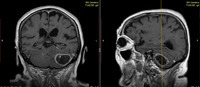
MRI Brain, Cerebellar neoplasm
1 of 10
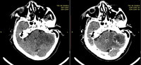
CT (brain), Cerebellar Neoplasm
2 of 10

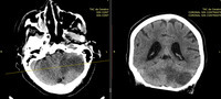
CT (brain), Cerebellar Neoplasm
3 of 10
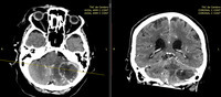
CT (brain), Cerebellar Neoplasm
4 of 10
CT (brain), Cerebellar Neoplasm
5 of 10
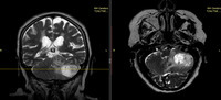
MRI Brain, Cerebellar Neoplasm
6 of 10
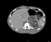
CT (abdomen), Splenic Rupture
7 of 10

CT (abdomen), Splenic Rupture
8 of 10
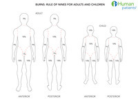
Burn Rule of Nine
9 of 10
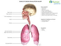
Signs of Potential Inhalation Injury
10 of 10
