Search
« Previous |
21 - 30 of 35
|
Next »
Search Results
Select an image to start the slideshow
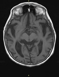
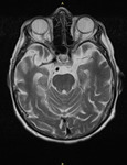
MRI Brain - Alzheimer's Disease
1 of 10
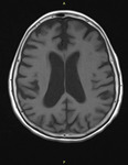
MRI Brain - Alzheimer's Disease
2 of 10
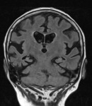
MRI Brain - Alzheimer's Disease
3 of 10
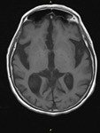
MRI Brain - Alzheimer's Disease
4 of 10
MRI Brain - Alzheimer's Disease
5 of 10
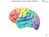
Functional Areas Of The Brain, Matching
6 of 10
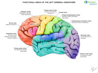
Functional Areas Of The Brain, Labeled
7 of 10
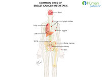
Breast Cancer Metastases
8 of 10
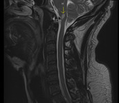
Lateral Medulla Infarct, MRI Brain
9 of 10
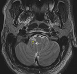
Lateral Medulla Infarct, MRI Brain
10 of 10
