Search
« Previous |
291 - 300 of 1,082
|
Next »
Search Results
Select an image to start the slideshow
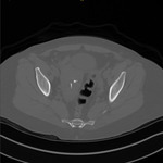
CT (abdomen), Acute Diverticulitis, Female
1 of 10
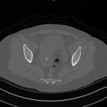
CT (abdomen), Acute Diverticulitis, Female
2 of 10
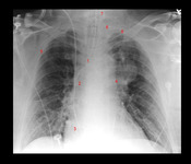
X-ray (chest), PA, Aortic Dissection, Adult Male
3 of 10
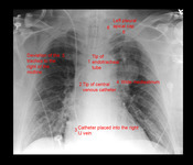
X-ray (chest), PA, Aortic Dissection, Adult Male
4 of 10
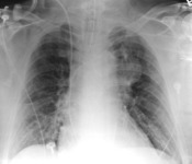
X-ray (chest), PA, Aortic Dissection, Adult Male
5 of 10
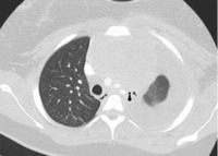
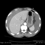
CT (chest), Lung Mass with Effusion and Left Deviation of the Trachea
6 of 10
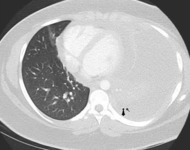
CT (chest), Lung Mass with Effusion and Left Deviation of the Trachea
7 of 10
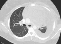
CT (chest), Lung Mass with Effusion and Left Deviation of the Trachea
8 of 10
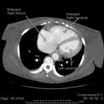
CT (chest), Right Heart Hypertrophy (annotated)
9 of 10
CT (abdomen), Hemoperitoneum
10 of 10
