Search
« Previous |
1 - 10 of 11
|
Next »
Search Results
Select an image to start the slideshow
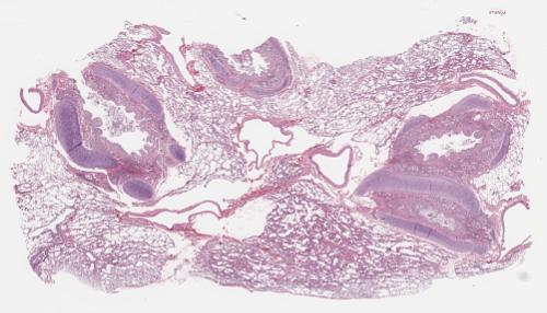
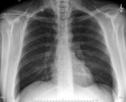
Lung, Asthma
1 of 10
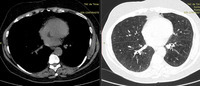
CT (chest), Chronic Obstructive Pulmonary Disease and Emphysema
2 of 10
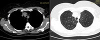
CT (chest), Chronic Obstructive Pulmonary Disease and Emphysema
3 of 10
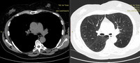
CT (chest), Chronic Obstructive Pulmonary Disease and Emphysema
4 of 10
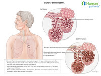
Chronic obstructive pulmonary disease (COPD) / Emphysema
5 of 10
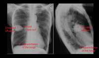
X-ray (chest), PA and Lateral, Encysted Effusion with Answers, Adult Male
6 of 10
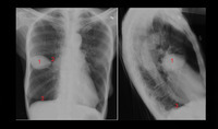
X-ray (chest), PA and Lateral, Encysted Effusion with Numbers, Adult Male
7 of 10
X-ray (chest), PA and Lateral, Encysted Effusion, Adult Male
8 of 10
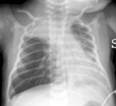
X-ray (chest), PA, Bronchiolitis, Infant
9 of 10
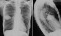
X-ray (chest), AP, Asthma with Pneumomediastinum, Adult Male
10 of 10
