Search
« Previous |
651 - 660 of 775
|
Next »
Search Results
Select an image to start the slideshow
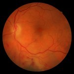
Branch Retinal Vein Occlusion, Funduscopic Exam
1 of 10

Diabetic Retinopathy, Funduscopic Exam
2 of 10
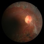
Diabetic Retinopathy, Funduscopic Exam
3 of 10
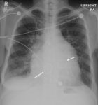
X-ray (chest), PA, Mitral Stenosis with Prosthetic Mitral and Aortic Valves, Adult Male
4 of 10
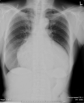
X-ray (chest), PA, Pericardial Effusion, Adult Female
5 of 10
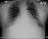
X-ray (chest), AP, Pleural Effusion and Congestive Heart Failure (CHF)
6 of 10
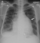
X-ray (chest), PA, Mitral Stenosis, Prosthetic Mitral/Aortic Valves, Adult Male
7 of 10
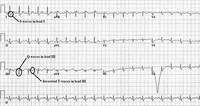
12-Lead ECG: right ventricular strain - with annotation
8 of 10
12-Lead ECG: right ventricular strain - with annotation
9 of 10
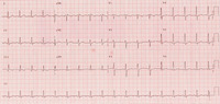
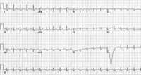
12-Lead ECG: right ventricular strain
10 of 10
