Search
« Previous |
1,391 - 1,400 of 2,694
|
Next »
Search Results
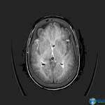
- Title:
- CT (brain), Subarachnoid Hemorrhage, Frontal Parietal Temporal Cortex, Sylvian Fissure
- Description:
- 4th ventricle tent Cerebelli
Frontal parietal temporal cortex
Sylvian fissure
- Keyword:
- X Ray Computerized Tomography, Tomography, Transmission Computed, Cine-CT, Hemorrhage, Subarachnoid, Perinatal Subarachnoid Hemorrhage, Computed Tomography, X-Ray, CAT Scan, X-Ray, X-Ray Computerized Axial Tomography, X-Ray Computer Assisted Tomography, CT Scan, X-Ray, SAH (Subarachnoid Hemorrhage), Tomography, X Ray Computed, X-Ray Tomography, Computed, Tomography, X-Ray Computer Assisted, Computerized Tomography, X Ray, Subarachnoid Hemorrhage, Intracranial, Tomography, X-Ray Computerized Axial, Subarachnoid Hemorrhage, Spontaneous, CT X Ray, Electron Beam Tomography, CAT Scan, X Ray, Subarachnoid Hemorrhage, Aneurysmal, Computed X Ray Tomography, Tomodensitometry, Tomography, Xray Computed, Computerized Tomography, X-Ray, Tomography, X-Ray Computerized, Electron Beam Computed Tomography, Diagnosis, X Ray Tomography, Computed
- Subject:
- Intracranial Hemorrhages, Multimodal Imaging, Subarachnoid Hemorrhage, Pathological Conditions, Signs and Symptoms, Tomography, X-Ray Computed, Diagnostic Techniques and Procedures, Pathologic Processes, Hemorrhage, Diagnostic Imaging
- Creator:
- i-Human Patients
- Publisher:
- i-Human Patients, Inc., Inc.
- Language:
- English
- Copyright Holder:
- i-Human Patients, Inc.
- Rights:
- http://www.i-human.com/service-agreement-print
- Resource Type:
- Medical Imaging
- Identifier:
- 1508
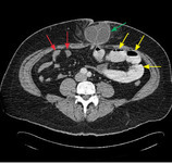
- Title:
- CT (abdomen), (axial), Small Bowel Obstruction (annotated)
- Description:
- Axial computed tomography scan showing dilated, contrast-filled loops of bowel on the patient's left (yellow arrows), with decompressed distal small bowel on the patient's right (red arrows)> The cause of obstruction, an incarcerated umbilical hernia, can also be seen (green arrow), with proximally dilated bowel entering the hernia and decompressed bowel existing the hernia.
- Keyword:
- X-Ray Computer Assisted Tomography, SBO, Obstruction, X Ray Tomography, Computed, X-Ray Tomography, Computed, Electron Beam Computed Tomography, Tomography, Transmission Computed, Electron Beam Tomography, Bowel, X-Ray Computerized Axial Tomography, Computed X Ray Tomography, X Ray Computerized Tomography, Tomography, X Ray Computed, Gastrointestinal Diseases, CAT Scan, X-Ray, Tomography, X-Ray Computerized, CT X Ray, Tomodensitometry, Intestinal Diseases, Computerized Tomography, X Ray, CAT Scan, X Ray, Diagnosis, Intestinal Obstruction, Tomography, Xray Computed, Tomography, X-Ray Computerized Axial, Computerized Tomography, X-Ray, CT Scan, X-Ray, Cine-CT, Computed Tomography, X-Ray, Tomography, X-Ray Computer Assisted, Small Bowel Obstruction, Digestive System Diseases
- Subject:
- Intestinal Diseases, Intestinal Obstruction, Tomography, X-Ray Computed, Digestive System Diseases, Multimodal Imaging, Diagnostic Techniques and Procedures, Gastrointestinal Diseases, Diagnostic Imaging
- Creator:
- Permission from American Family Physician
- Publisher:
- i-Human Patients, Inc.
- Language:
- English
- Copyright Holder:
- American Family Physician
- Rights:
- http://www.i-human.com/service-agreement-print
- Resource Type:
- Medical Imaging
- Identifier:
- 1504
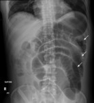
- Title:
- CT (abdomen), (lateral supine), Small Bowel Obstruction (annotated)
- Description:
- Supine view of the abdomen in a patient with intestinal obstruction. Dilated loops of small bowel are visible (arrows).
- Keyword:
- Intestinal Obstruction, X Ray Computerized Tomography, Computed Tomography, X-Ray, Tomodensitometry, Electron Beam Tomography, Tomography, X-Ray Computer Assisted, Tomography, X-Ray Computerized, X-Ray Computerized Axial Tomography, CT Scan, X-Ray, Electron Beam Computed Tomography, X-Ray Tomography, Computed, Tomography, X-Ray Computerized Axial, Computerized Tomography, X Ray, CAT Scan, X-Ray, X Ray Tomography, Computed, Gastrointestinal Diseases, X-Ray Computer Assisted Tomography, Tomography, X Ray Computed, Digestive System Diseases, Tomography, Transmission Computed, Small Bowel Obstruction, Bowel, Diagnosis, CT X Ray, Computerized Tomography, X-Ray, Intestinal Diseases, Cine-CT, Computed X Ray Tomography, Obstruction, SBO, Tomography, Xray Computed, CAT Scan, X Ray
- Subject:
- Gastrointestinal Diseases, Intestinal Diseases, Multimodal Imaging, Intestinal Obstruction, Tomography, X-Ray Computed, Diagnostic Imaging, Diagnostic Techniques and Procedures, Digestive System Diseases
- Creator:
- American Family Physician
- Publisher:
- i-Human Patients, Inc.
- Language:
- English
- Copyright Holder:
- American Family Physician
- Rights:
- http://www.i-human.com/service-agreement-print
- Resource Type:
- Medical Imaging
- Identifier:
- 1503
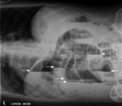
- Title:
- CT (abdomen), (lateral decubitus), Small Bowel Obstruction (annotated)
- Description:
- Lateral decubitus view of the abdomen, showing air-fluid levels consistent with intestinal obstruction (arrows).
- Keyword:
- Tomodensitometry, X-Ray Tomography, Computed, Gastrointestinal Diseases, Tomography, X-Ray Computer Assisted, Bowel, Tomography, X-Ray Computerized, CAT Scan, X Ray, Tomography, X-Ray Computerized Axial, Obstruction, Tomography, X Ray Computed, X-Ray Computer Assisted Tomography, CT Scan, X-Ray, Diagnosis, X-Ray Computerized Axial Tomography, CT X Ray, Tomography, Xray Computed, Computed X Ray Tomography, Electron Beam Computed Tomography, Intestinal Diseases, Tomography, Transmission Computed, Cine-CT, Small Bowel Obstruction, Digestive System Diseases, SBO, X Ray Tomography, Computed, CAT Scan, X-Ray, Computerized Tomography, X Ray, Computed Tomography, X-Ray, X Ray Computerized Tomography, Intestinal Obstruction, Computerized Tomography, X-Ray, Electron Beam Tomography
- Subject:
- Multimodal Imaging, Diagnostic Imaging, Intestinal Diseases, Digestive System Diseases, Diagnostic Techniques and Procedures, Gastrointestinal Diseases, Intestinal Obstruction, Tomography, X-Ray Computed
- Creator:
- American Family Physician
- Publisher:
- i-Human Patients, Inc.
- Language:
- English
- Copyright Holder:
- American Family Physician
- Rights:
- http://www.i-human.com/service-agreement-print
- Identifier:
- 1502
- Title:
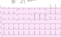
- 12-Lead ECG: Normal Sinus Rhythm (NSR) ~69 bpm
- Description:
- 12-Lead ECG: NSR 69 bpm
- Keyword:
- Pulse Rate, Electrocardiogram, Heart, Heart Rate Control, Chronotropism, Cardiac, Rates, Pulse, Chronotropy, Cardiac, Electrocardiograph, EKG, Cardiac Chronotropy, ECG, Diagnosis, Cardiac, Pulse Rates, Chronotropism, Rates, Heart, Rate, Heart, Heart Rates, Rate, Pulse, Rate Control, Heart, Control, Heart Rate
- Subject:
- Diagnostic Techniques and Procedures, Hemodynamics, Electrocardiography, Heart Rate, Vital Signs, Diagnostic Techniques, Cardiovascular, Physical Examination
- Creator:
- Avery Ellis, MD, PhD.University of Buffalo School of Medicine
- Publisher:
- University of Buffalo School of Medicine
- Language:
- English
- Copyright Holder:
- University at Buffalo School of Medicine
- Rights:
- http://www.i-human.com/service-agreement-print
- Resource Type:
- Chart/Diagram
- Identifier:
- 1500

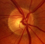
- Title:
- Optic Disc, Edema, Funduscopic Exam
- Description:
- Funduscopic - Optic disc, edema
- Keyword:
- Papilledema, Diagnostic Techniques, Ophthalmological, Diagnosis
- Subject:
- Diagnostic Techniques and Procedures, Papilledema, Eye Diseases, Optic Nerve Diseases, Nervous System Diseases, Ophthalmoscopy
- Publisher:
- Flylib.com
- Language:
- English
- Copyright Holder:
- Flylib.com
- Rights:
- http://www.i-human.com/service-agreement-print
- Resource Type:
- Photo
- Identifier:
- 1497
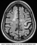
- Title:
- MRI, Brain - Multiple Sclerosis
- Description:
- MRI: Brain multiple sclerosis
- Keyword:
- Magnetization Transfer Contrast Imaging, MR Tomography, Imaging, Chemical Shift, fMRI, Diagnosis, MRI Scans, NMR Tomography, MS, Tomography, Proton Spin, Tomography, NMR, Anatomy, Cross-Sectional, Magnetic Resonance Imaging, Functional, Demyelinating Autoimmune Diseases, CNS, NMR Imaging, Zeugmatography, Functional Magnetic Resonance Imaging, Tomography, MR, Proton Spin Tomography, Magnetic Resonance Spectroscopy, Multiple Sclerosis, Acute Fulminating, Chemical Shift Imaging, Sclerosis, Disseminated, MRI, Functional
- Subject:
- Multimodal Imaging, Nervous System Diseases, Multiple Sclerosis, Diagnostic Techniques and Procedures, Diagnostic Imaging, Magnetic Resonance Imaging, Autoimmune Diseases of the Nervous System
- Creator:
- Rush Medical College
- Publisher:
- Rush Medical College
- Language:
- English
- Copyright Holder:
- Rush Medical College
- Rights:
- http://www.i-human.com/service-agreement-print
- Identifier:
- 1495
- Title:
- Optic Disc, Normal OD, Fundoscopic Exam
- Description:
- Funduscopic - Optic Disc, Normal OD
- Keyword:
- Diagnosis, Eye, Diagnostic Techniques, Ophthalmological, eye exam
- Subject:
- Ophthalmoscopy, Sense Organs, Diagnostic Techniques and Procedures, Retina, Eye
- Creator:
- Hong Kong Medical Association
- Publisher:
- i-Human Patients, Inc.
- Language:
- English
- Copyright Holder:
- Hong Kong Medical Association
- Rights:
- http://www.i-human.com/service-agreement-print
- Resource Type:
- Photo
- Identifier:
- 1496
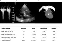
- Title:
- Transthoracic Echocardiogram (TTE), Aortic Stenosis with Valve Gradients
- Description:
- Echocardiogram - Aortic Stenosis (still) with valve gradients
- Keyword:
- M-Mode Echocardiography, 2D Echocardiography, Echocardiography, Contrast, Echocardiography, 2D, Two-Dimensional Echocardiography, aorta, aortic, stenosis, stricture, pathological constriction,, Echocardiography, Cross-Sectional, Transthoracic Echocardiography, Echocardiography, Transthoracic, Contrast Echocardiography, Echocardiography, Two-Dimensional, Echocardiography, M-Mode, Echocardiography, 2-D, Cross-Sectional Echocardiography, Diagnosis, 2-D Echocardiography
- Subject:
- Echocardiography, Aorta, Diagnostic Techniques and Procedures, Arteries, Cardiovascular System, Diagnostic Imaging, Pathological Conditions, Anatomical, Constriction, Pathologic, Multimodal Imaging, Cardiac Imaging Techniques, Blood Vessels, Pathological Conditions, Signs and Symptoms
- Creator:
- Jah Won Koo, MD
- Publisher:
- i-Human Patients, Inc.
- Language:
- English
- Copyright Holder:
- Jah Won Koo, MD
- Rights:
- http://www.i-human.com/service-agreement-print
- Resource Type:
- Medical Imaging
- Identifier:
- 1476
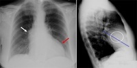
- Title:
- X-ray (chest PA), Aortic Stenosis with Annotation
- Description:
- CXR PA and Lateral - Adult male, aortic stenosis with annotation
- Keyword:
- Radiology, Diagnostic X-Ray, Roentgenography, Radiography, Diagnostic X-Ray, Diagnostic X-Ray Radiology, Diagnosis, X-Ray Radiology, Diagnostic, X-Ray, Diagnostic, aorta, aortic, stenosis, stricture, pathological constriction,
- Subject:
- Diagnostic Techniques and Procedures, Multimodal Imaging, Blood Vessels, Cardiovascular System, Diagnostic Imaging, Aorta, Arteries
- Creator:
- i-Human Patients, Inc., Inc.
- Publisher:
- i-Human Patients, Inc., Inc.
- Language:
- English
- Copyright Holder:
- i-Human Patients, Inc., Inc.
- Rights:
- http://www.i-human.com/service-agreement-print
- Identifier:
- 1475
