Search

You searched for: Keyword Diagnosis Remove constraint Keyword: Diagnosis Subject Pathological Conditions, Signs and Symptoms Remove constraint Subject: Pathological Conditions, Signs and Symptoms

Search Results

  1. CT (chest), Pulmonary Embolism with Infarction, Superior to Inferior

    show more
    Description: CT Chest - Pulmonary embolism with infarct superior to inferior
    Keywords: CAT Scan, X-Ray, Tomography, X-Ray Computerized, Electron Beam Computed Tomography, Tomography, X-Ray Computer Assisted, X-Ray Computer Assisted Tomography, X-Ray Computerized Axial Tomography, X-Ray Tomography, Computed, CAT Scan, X Ray, CT X Ray, Pulmonary Thromboembolism, X Ray Tomography, Computed, Pulmonary Embolisms, Embolism, Pulmonary, Pulmonary Thromboembolisms, Tomography, X-Ray Computerized Axial, Tomography, Transmission Computed, Computerized Tomography, X Ray, Infarct, Tomography, X Ray Computed, Embolisms, Pulmonary, Cine-CT, X Ray Computerized Tomography, Thromboembolisms, Pulmonary, CT Scan, X-Ray, Computerized Tomography, X-Ray, Diagnosis, Computed X Ray Tomography, Tomography, Xray Computed, Thromboembolism, Pulmonary, Electron Beam Tomography, Computed Tomography, X-Ray, Tomodensitometry
  2. Transthoracic Echocardiogram (TTE), Aortic Stenosis (SAX through AoV)

    show more
    Description: Echocardiogram - Aortic Stenosis (Short Axis through AoV)
    Keywords: 2-D Echocardiography, Echocardiography, M-Mode, Echocardiography, Cross-Sectional, M-Mode Echocardiography, Cross-Sectional Echocardiography, Echocardiography, Transthoracic, Diagnosis, Echocardiography, Contrast, Contrast Echocardiography, Echocardiography, 2D, Echocardiography, Two-Dimensional, Two-Dimensional Echocardiography, aorta, aortic, stenosis, stricture, pathological constriction,, Transthoracic Echocardiography, Echocardiography, 2-D, 2D Echocardiography
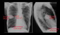
  3. X-ray (chest), PA and Lateral, Encysted Effusion with Answers, Adult Male

    show more
    Description: 1. Fluid density (same density as the heart), biconcave, lens shape consistent with an encysted effusion in the horizontal fissure. 2. Fluid "tails out" along the horizontal fissure characteristic of an encysted effusion 3. Note the hyperinflation of the lungs (more than 10 rib spaces visible) and the flattening of the usual dome-shaped configuration of the diaphragms, also visible on the lateral view. This is consistent with "air trapping" conditions such as asthma and emphysema.
    Keywords: Diagnosis, Radiology, Diagnostic X-Ray, Diagnostic X-Ray Radiology, Bronchial Asthma, lung effusion, Roentgenography, X-Ray, Diagnostic, Diagnostic X-Ray, Asthma, Bronchial, Radiography, X-Ray Radiology, Diagnostic
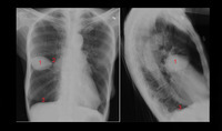
  4. X-ray (chest), PA and Lateral, Encysted Effusion with Numbers, Adult Male

    show more
    Description: 1. Fluid density (same density as the heart), biconcave, lens shape consistent with an encysted effusion in the horizontal fissure. 2. Fluid "tails out" along the horizontal fissure characteristic of an encysted effusion 3. Note the hyperinflation of the lungs (more than 10 rib spaces visible) and the flattening of the usual dome-shaped configuration of the diaphragms, also visible on the lateral view. This is consistent with "air trapping" conditions such as asthma and emphysema.
    Keywords: Radiography, Asthma, Bronchial, Radiology, Diagnostic X-Ray, X-Ray Radiology, Diagnostic, Diagnostic X-Ray Radiology, Bronchial Asthma, Diagnostic X-Ray, Roentgenography, lung effusion, X-Ray, Diagnostic, Diagnosis
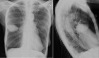
  5. X-ray (chest), PA and Lateral, Encysted Effusion, Adult Male

    show more
    Description: 1. Fluid density (same density as the heart), biconcave, lens shape consistent with an encysted effusion in the horizontal fissure. 2. Fluid "tails out" along the horizontal fissure characteristic of an encysted effusion 3. Note the hyperinflation of the lungs (more than 10 rib spaces visible) and the flattening of the usual dome-shaped configuration of the diaphragms, also visible on the lateral view. This is consistent with "air trapping" conditions such as asthma and emphysema.
    Keywords: X-Ray, Diagnostic, Roentgenography, Asthma, Bronchial, Diagnostic X-Ray Radiology, Bronchial Asthma, Diagnostic X-Ray, Radiography, lung effusion, X-Ray Radiology, Diagnostic, Diagnosis, Radiology, Diagnostic X-Ray
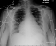
  6. X-ray (chest), AP, Progressive Right Heart Hypertrophy

    show more
    Description: CXR AP - Adult female, baseline and progressive right heart hypertrophy
    Keywords: Radiography, Diagnostic X-Ray, Roentgenography, Diagnosis, Diagnostic X-Ray Radiology, X-Ray Radiology, Diagnostic, X-Ray, Diagnostic, Radiology, Diagnostic X-Ray, Progressive Right Heart Hypertrophy, Heart, Hypertrophy
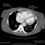
  7. CT (chest), Right Heart Hypertrophy (annotated)

    show more
    Description: CT - Chest - right heart hypertrophy with and without labels
    Keywords: Tomography, X-Ray Computer Assisted, Hypertrophy, Tomography, Xray Computed, X-Ray Computer Assisted Tomography, CT X Ray, CAT Scan, X Ray, Electron Beam Computed Tomography, Tomography, X-Ray Computerized, Tomography, X-Ray Computerized Axial, Electron Beam Tomography, Cine-CT, Heart, Tomography, Transmission Computed, Tomodensitometry, Computerized Tomography, X Ray, Diagnosis, CAT Scan, X-Ray, CT Scan, X-Ray, X Ray Tomography, Computed, Computed X Ray Tomography, X-Ray Computerized Axial Tomography, Computed Tomography, X-Ray, Tomography, X Ray Computed, Computerized Tomography, X-Ray, X-Ray Tomography, Computed, X Ray Computerized Tomography, Right Heart Hypertrophy
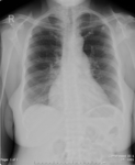
  8. X-ray (chest), AP, Baseline Right Heart Hypertrophy

    show more
    Description: CXR AP - Adult female, baseline and progressive right heart hypertrophy
    Keywords: Radiography, Diagnostic X-Ray Radiology, Roentgenography, Radiology, Diagnostic X-Ray, Diagnosis, Diagnostic X-Ray, X-Ray, Diagnostic, Hypertrophy, X-Ray Radiology, Diagnostic, Right Heart Hypertrophy
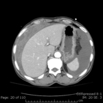
  9. CT (abdomen), Hemoperitoneum

    show more
    Description: Emphysema with no infiltrate or pneumothorax. The visualized portion of the heart is normal in size. There are no pleural or pericardial effusions. There are coronary artery calcifications. There is active arterial extravasation from a peripheral liver mass at the dome of the liver, which measures approximately 3.8 x 3.2 cm. This mass enhances centrally and peripherally during the arterial phase of this examination. There is extensive hemoperitoneum, in varying stages of density. When compared with the prior CT scan, there was a lesion within the liver which demonstrated early arterial . The liver contour is smooth. There is a second less well-defined area of increased enhancement (series 2 image 81) of uncertain etiology. There are 2 hypodensities in the right hepatic lobe which are too small to characterize. There are small periportal and paraaortic lymph nodes. The spleen and pancreas are normal in appearance. The adrenal glands are unremarkable. There are multiple radiopaque gallstones. The kidneys enhance normally and symmetrically. There is a 2.1 cm renal cyst within the left interpolar region. There are multiple other renal hypodensities which are too small to characterize, however probably represents cysts. There is no hydronephrosis. The bowel is normal in caliber without evidence of bowel wall thickening. There are scattered colonic diverticula. The aorta and its visceral branches are patent. There are postsurgical changes from recent aortobifemoral graft placement with patency of the graft. The pelvic viscera are unremarkable. There are no fractures. There are mild degenerative changes of the spine. IMPRESSION: Active extravasation with extensive hemoperitoneum arising from a 3.8 cm peripherally enhancing lesion within the dome of the liver.
    Keywords: Tomography, X-Ray Computerized Axial, CAT Scan, X-Ray, Diagnosis, CAT Scan, X Ray, Computerized Tomography, X Ray, Tomography, X-Ray Computerized, Computed X Ray Tomography, Electron Beam Tomography, X Ray Tomography, Computed, CT Scan, X-Ray, X Ray Computerized Tomography, Tomography, X-Ray Computer Assisted, X-Ray Computer Assisted Tomography, Tomography, Transmission Computed, Computerized Tomography, X-Ray, X-Ray Tomography, Computed, Tomodensitometry, Tomography, X Ray Computed, air in lungs, Computed Tomography, X-Ray, CT X Ray, Cine-CT, Electron Beam Computed Tomography, Tomography, Xray Computed, X-Ray Computerized Axial Tomography
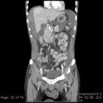
  10. CT (abdomen), (coronal), Hemoperitoneum

    show more
    Description: Emphysema with no infiltrate or pneumothorax. The visualized portion of the heart is normal in size. There are no pleural or pericardial effusions. There are coronary artery calcifications. There is active arterial extravasation from a peripheral liver mass at the dome of the liver, which measures approximately 3.8 x 3.2 cm. This mass enhances centrally and peripherally during the arterial phase of this examination. There is extensive hemoperitoneum, in varying stages of density. When compared with the prior CT scan, there was a lesion within the liver which demonstrated early arterial . The liver contour is smooth. There is a second less well-defined area of increased enhancement (series 2 image 81) of uncertain etiology. There are 2 hypodensities in the right hepatic lobe which are too small to characterize. There are small periportal and paraaortic lymph nodes. The spleen and pancreas are normal in appearance. The adrenal glands are unremarkable. There are multiple radiopaque gallstones. The kidneys enhance normally and symmetrically. There is a 2.1 cm renal cyst within the left interpolar region. There are multiple other renal hypodensities which are too small to characterize, however probably represents cysts. There is no hydronephrosis. The bowel is normal in caliber without evidence of bowel wall thickening. There are scattered colonic diverticula. The aorta and its visceral branches are patent. There are postsurgical changes from recent aortobifemoral graft placement with patency of the graft. The pelvic viscera are unremarkable. There are no fractures. There are mild degenerative changes of the spine. IMPRESSION: Active extravasation with extensive hemoperitoneum arising from a 3.8 cm peripherally enhancing lesion within the dome of the liver.
    Keywords: X-Ray Computerized Axial Tomography, Electron Beam Tomography, Tomography, X-Ray Computerized, Tomodensitometry, Tomography, Transmission Computed, CAT Scan, X-Ray, Cine-CT, Computed Tomography, X-Ray, Electron Beam Computed Tomography, Tomography, Xray Computed, Diagnosis, X Ray Tomography, Computed, X-Ray Tomography, Computed, CAT Scan, X Ray, X-Ray Computer Assisted Tomography, CT X Ray, X Ray Computerized Tomography, Tomography, X-Ray Computerized Axial, Tomography, X Ray Computed, air in lungs, Tomography, X-Ray Computer Assisted, Computerized Tomography, X Ray, Computed X Ray Tomography, Computerized Tomography, X-Ray, CT Scan, X-Ray, CT Scan, Coronal Plane