Search
« Previous |
201 - 210 of 2,770
|
Next »
Search Results

- Title:
- X-ray (chest), AP Lat - Pleural Effusion
- Description:
- There is a large opacity in the right lung base consistent with a large right pleural effusion. This was not present at the time of the comparison radiograph.
The radiographic appearance of the chest is otherwise unremarkable. The heart is normal in size. The aerated portions of both lungs appear normal. The left hemidiaphragm is well-defined, as is the left costophrenic sulcus.
IMPRESSION: New, large, right pleural effusion.
- Keyword:
- Computer Echotomography, Ultrasonic Imaging, Ultrasonic Tomography, Diagnosis, Ultrasonic, Echography, Tomography, Ultrasonic, Echotomography, Computer, Effusion, Ultrasonic Diagnosis, Ultrasound Imaging, Sonography, Medical, Pleura, Echotomography, Diagnosis
- Subject:
- Multimodal Imaging, Diagnostic Imaging, Pleural Effusion, Diagnostic Techniques and Procedures, Ultrasonography
- Creator:
- Rush Medical College
- Publisher:
- Rush Medical College
- Language:
- English
- Rights:
- http://www.i-human.com/service-agreement-print
- Resource Type:
- Medical Imaging
- Identifier:
- 3223
- Title:
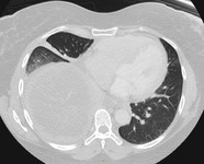
- CT (chest), Pleural Effusion
- Description:
- Demonstrates associated pleural effusion and pleural thickening at the posterior aspect of the R lung (compare to healthy left lung). Note the part of the mass anterior to the effusion (hyperdense relative to the effusion).
- Keyword:
- Tomography, Ultrasonic, Echotomography, Echotomography, Computer, Computer Echotomography, Sonography, Medical, Ultrasonic Tomography, Echography, Pleura, Diagnosis, Ultrasonic Diagnosis, Ultrasonic Imaging, Ultrasound Imaging, Effusion, Diagnosis, Ultrasonic
- Subject:
- Diagnostic Techniques and Procedures, Diagnostic Imaging, Pleural Effusion, Ultrasonography, Multimodal Imaging
- Creator:
- Rush Medical College
- Publisher:
- Rush Medical College
- Language:
- English
- Rights:
- http://www.i-human.com/service-agreement-print
- Resource Type:
- Medical Imaging
- Identifier:
- 3225
- Title:
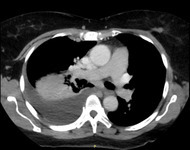
- CT (chest), Pleural Effusion
- Description:
- Demonstrates associated pleural effusion and pleural thickening at the posterior aspect of the R lung (compare to healthy left lung). Note the part of the mass anterior to the effusion (hyperdense relative to the effusion).
- Keyword:
- Effusion, Ultrasonic Diagnosis, Ultrasound Imaging, Diagnosis, Echotomography, Tomography, Ultrasonic, Echography, Sonography, Medical, Echotomography, Computer, Diagnosis, Ultrasonic, Pleura, Ultrasonic Tomography, Computer Echotomography, Ultrasonic Imaging
- Subject:
- Multimodal Imaging, Diagnostic Imaging, Diagnostic Techniques and Procedures, Pleural Effusion, Ultrasonography
- Creator:
- Rush Medical College
- Publisher:
- Rush Medical College
- Language:
- English
- Rights:
- http://www.i-human.com/service-agreement-print
- Resource Type:
- Medical Imaging
- Identifier:
- 3225
- Title:
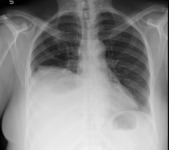
- X-ray (chest), AP Lat - Pleural Effusion
- Description:
- There is a large opacity in the right lung base consistent with a large right pleural effusion. This was not present at the time of the comparison radiograph.
The radiographic appearance of the chest is otherwise unremarkable. The heart is normal in size. The aerated portions of both lungs appear normal. The left hemidiaphragm is well-defined, as is the left costophrenic sulcus.
IMPRESSION: New, large, right pleural effusion.
- Keyword:
- Ultrasonic Tomography, Sonography, Medical, Diagnosis, Ultrasonic, Diagnosis, Echography, Echotomography, Computer, Computer Echotomography, Tomography, Ultrasonic, Pleura, Effusion, Ultrasonic Diagnosis, Echotomography, Ultrasonic Imaging, Ultrasound Imaging
- Subject:
- Ultrasonography, Diagnostic Techniques and Procedures, Pleural Effusion, Diagnostic Imaging, Multimodal Imaging
- Creator:
- Rush Medical College
- Publisher:
- Rush Medical College
- Language:
- English
- Rights:
- http://www.i-human.com/service-agreement-print
- Resource Type:
- Medical Imaging
- Identifier:
- 3223
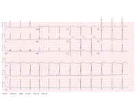
- Title:
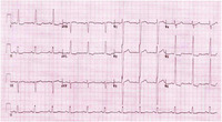
- 12-Lead ECG: Left ventricular hypertrophy (LV)
- Description:
- Rate: 70 bpm
- Keyword:
- Aortic Stenosis, Ventricular Hypertrophy, Left, Left Ventricular Hypertrophies, ECG, Hypertension, Electrocardiogram, Electrocardiograph, Ventricular Hypertrophies, Left, Heart Ventricles, EKG, Heart, Diagnosis, Left Ventricular Hypertrophy, Hypertrophies, Left Ventricular
- Subject:
- Heart Diseases, Diagnostic Techniques, Cardiovascular, Tachycardia, Pathological Conditions, Anatomical, Cardiomegaly, Diagnostic Techniques and Procedures, Hypertrophy, Left Ventricular, Electrocardiography
- Creator:
- Brian Neubauer, MD, FACP, Major, USAF, MC
Assistant Professor of Medicine Cardiopulmonary-renal Module Director
F. Edward Hebert School of Medicine Uniformed Services University of the Health Sciences
- Publisher:
- Brian Neubauer, MD, FACP, Major, USAF, MC
Assistant Professor of Medicine Cardiopulmonary-renal Module Director
F. Edward Hebert School of Medicine Uniformed Services University of the Health Sciences
- Language:
- English
- Rights:
- http://www.i-human.com/service-agreement-print
- Resource Type:
- Chart/Diagram
- Identifier:
- 3221

- Title:
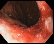
- Endoscopy, Mallory-Weiss tear
- Description:
- Picture from endoscopy showing Mallory-Weiss tear
- Keyword:
- colonoscope, Intestine, Large, Minimally Invasive Surgical Procedures, Colonoscopy, Medical procedure, Diagnosis, Digestive System Surgical Procedures
- Subject:
- Diagnostic Techniques and Procedures, Intestines, Colonoscopy, Endoscopy, Gastrointestinal, Endoscopy, Digestive System, Endoscopy, Diagnostic Techniques, Surgical, Surgical Procedures, Operative, Diagnostic Techniques, Digestive System, Mallory-Weiss Syndrome, Colon
- Creator:
- Joseph J. Rencic, MD, FACP Associate Professor of Internal MedicineTufts Medical CenterTufts University School of Medicine
- Contributor:
- Tufts University School of Medicine
- Publisher:
- Joseph J. Rencic, MD, FACP Associate Professor of Internal MedicineTufts Medical CenterTufts University School of Medicine
- Language:
- English
- Rights:
- http://www.i-human.com/service-agreement-print
- Identifier:
- 3217
- Title:
- Endoscopy, Mallory-Weiss tear
- Description:
- Picture from endoscopy showing Mallory-Weiss tear
- Keyword:
- colonoscope, Minimally Invasive Surgical Procedures, Colonoscopy, Medical procedure, Digestive System Surgical Procedures, Intestine, Large, Diagnosis
- Subject:
- Mallory-Weiss Syndrome, Diagnostic Techniques, Surgical, Colonoscopy, Endoscopy, Endoscopy, Gastrointestinal, Colon, Endoscopy, Digestive System, Diagnostic Techniques, Digestive System, Diagnostic Techniques and Procedures, Intestines, Surgical Procedures, Operative
- Creator:
- Joseph J. Rencic, MD, FACP Associate Professor of Internal MedicineTufts Medical CenterTufts University School of Medicine
- Contributor:
- Tufts Medical Center
- Publisher:
- Joseph J. Rencic, MD, FACP Associate Professor of Internal MedicineTufts Medical CenterTufts University School of Medicine
- Language:
- English
- Rights:
- http://www.i-human.com/service-agreement-print
- Identifier:
- 3217
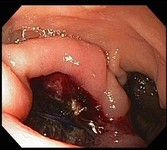
- Title:
- Endoscopy - Aortoenteric Fistula
- Description:
- Aortoenteric fistula
- Keyword:
- endoscopy, Minimally Invasive Surgical Procedures, Diagnosis
- Subject:
- Surgical Procedures, Operative, Aorta, Endoscopy, Biopsy, Diagnostic Techniques and Procedures, Diagnostic Techniques, Surgical
- Creator:
- Mike Sullivan MD Tufts Medical Center/ Tufts University School of Medicine
- Contributor:
- Tufts Medical Center
- Publisher:
- Mike Sullivan MD Tufts Medical Center/ Tufts University School of Medicine
- Language:
- English
- Rights:
- http://www.i-human.com/service-agreement-print
- Resource Type:
- Photo
- Identifier:
- 3215
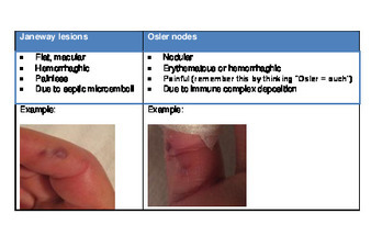
- Title:
- Compare Janeway and Osler
- Description:
- Janeway lesions vs Osler nodes
- Keyword:
- patient diagnosis, diagnostic reasoning, patient evaluation, patient symptoms, clinical reasoning skills, Diagnosis
- Subject:
- Diagnostic Techniques and Procedures
- Creator:
- Jennifer Babik MD
- Publisher:
- Jennifer Babik MD
- Language:
- English
- Rights:
- http://www.i-human.com/service-agreement-print
- Resource Type:
- Photo
- Identifier:
- 3216
- Title:
- 12 Lead ECG:72 BPM, non-specific T wave anormalities
- Description:
- 72 BPM
QRS Axis 42 degrees
PR interval 154 ms
QTc 422 ms
NSR, with voltage criteria for left ventricular hypertrophy
Non-specific T wave abnormalities
- Keyword:
- Chronotropy, Cardiac, Rate, Heart, Artificial Cardiac Pacemakers, Cardiac Chronotropy, Heart Rates, Heart, Rate, Pulse, Heart Rate Control, Pulse Rates, Diagnosis, Pulse Rate, Electrocardiogram, Pacemaker, Artificial, Control, Heart Rate, Rates, Heart, Paced Rhythm, Artificial Pacemaker, Cardiac, ECG, Chronotropism, Cardiac, Rates, Pulse, Rate Control, Heart, Chronotropism, Electrocardiograph, EKG
- Subject:
- Diagnostic Techniques and Procedures, Vital Signs, Diagnostic Techniques, Cardiovascular, Electrocardiography, Heart Rate, Hemodynamics, Physical Examination
- Creator:
- David Beiser, MD
- Publisher:
- David Beiser, MD
- Language:
- English
- Rights:
- http://www.i-human.com/service-agreement-print
- Resource Type:
- Chart/Diagram
- Identifier:
- 3211
