Search
« Previous |
1 - 10 of 61
|
Next »
Search Results
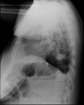
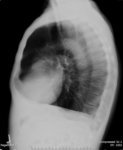
- Title:
- X-ray (chest Lateral), Granulomatosis with Polyangiitis (Wegeners Granulomatosis)
- Description:
- Wegeners granulomatosis
Adult female
Granulomatosis with polyangiitis (GPA)
Multiple b/l nodular opacities >5 cm in diam right mid-lung field and apices bilaterally; normal cor size; no effusion
- Keyword:
- Radiography, X-Ray, Diagnostic, Radiology, Diagnostic X-Ray, Wegener Granulomatosis, Diagnosis, Diagnostic X-Ray Radiology, Wegener's Granulomatosis, Diagnostic X-Ray, X-Ray Radiology, Diagnostic, Granulomatosis, Wegener's, Roentgenography
- Subject:
- Granulomatosis with Polyangiitis, Multimodal Imaging, Autoimmune Diseases, Diagnostic Imaging, Immune System Diseases, Anti-Neutrophil Cytoplasmic Antibody-Associated Vasculitis, Diagnostic Techniques and Procedures
- Creator:
- i-Human Patients, Inc., Inc.
- Publisher:
- i-Human Patients, Inc., Inc.
- Language:
- English
- Copyright Holder:
- i-Human Patients, Inc.
- Rights:
- http://www.i-human.com/service-agreement-print
- Identifier:
- 1721
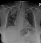
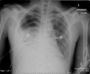
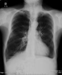
- Title:
- X-ray (chest PA), Granulomatosis with Polyangiitis (Wegeners Granulomatosis)
- Description:
- Wegeners granulomatosis
Adult Female, CXR PA
Granulomatosis with polyangiitis (GPA)
Multiple b/l nodular opacities >5 cm in diam right mid-lung field and apices bilaterally; normal cor size; no effusion
- Keyword:
- X-Ray Radiology, Diagnostic, Roentgenography, Wegener's Granulomatosis, Radiology, Diagnostic X-Ray, Granulomatosis, Wegener's, Wegener Granulomatosis, X-Ray, Diagnostic, Radiography, Diagnosis, Diagnostic X-Ray, Diagnostic X-Ray Radiology
- Subject:
- Anti-Neutrophil Cytoplasmic Antibody-Associated Vasculitis, Diagnostic Techniques and Procedures, Autoimmune Diseases, Immune System Diseases, Diagnostic Imaging, Granulomatosis with Polyangiitis, Multimodal Imaging
- Creator:
- i-Human Patients, Inc., Inc.
- Publisher:
- i-Human Patients, Inc., Inc.
- Language:
- English
- Copyright Holder:
- i-Human Patients, Inc.
- Rights:
- http://www.i-human.com/service-agreement-print
- Identifier:
- 1721
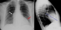
- Title:
- X-ray (chest PA), Aortic Stenosis with Annotation
- Description:
- CXR PA and Lateral - Adult male, aortic stenosis with annotation
- Keyword:
- Radiology, Diagnostic X-Ray, Roentgenography, Radiography, Diagnostic X-Ray, Diagnostic X-Ray Radiology, Diagnosis, X-Ray Radiology, Diagnostic, X-Ray, Diagnostic, aorta, aortic, stenosis, stricture, pathological constriction,
- Subject:
- Diagnostic Techniques and Procedures, Multimodal Imaging, Blood Vessels, Cardiovascular System, Diagnostic Imaging, Aorta, Arteries
- Creator:
- i-Human Patients, Inc., Inc.
- Publisher:
- i-Human Patients, Inc., Inc.
- Language:
- English
- Copyright Holder:
- i-Human Patients, Inc., Inc.
- Rights:
- http://www.i-human.com/service-agreement-print
- Identifier:
- 1475
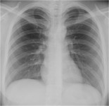
- Title:
- X-ray (chest), PA, Adult Male, Normal
- Description:
- CXR PA - Adult male, normal
- Keyword:
- Diagnostic X-Ray Radiology, Diagnostic X-Ray, Diagnosis, Thoraces, Chest, Roentgenography, X-Ray Radiology, Diagnostic, Radiography, X-Ray, Diagnostic, Radiology, Diagnostic X-Ray
- Subject:
- Diagnostic Techniques and Procedures, Thorax, Multimodal Imaging, Torso, Diagnostic Imaging, Body Regions
- Creator:
- Chris DeMauro, MD
- Publisher:
- i-Human Patients, Inc., Inc.
- Language:
- English
- Copyright Holder:
- Chris DeMauro, MD
- Rights:
- http://www.i-human.com/service-agreement-print
- Resource Type:
- Medical Imaging
- Identifier:
- 1469
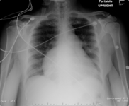
- Title:
- X-ray (chest), AP, Post-gunshot Wound, With and Without Chest Tube
- Description:
- Adult male status post-gunshot wound
There is a right hemothorax with a right apical pneumothorax. There is airspace disease at the right lung base. There is a small amount of subcutaneous emphysema overlying the lateral aspect of the right hemithorax. The left lung is well expanded and clear. The visualized portions of the cardiac, hilar, and mediastinal silhouettes are unremarkable. There is also a comminuted fracture of the left humerus.
- Keyword:
- X-Ray Radiology, Diagnostic, Roentgenography, Radiography, Diagnostic X-Ray Radiology, X-Ray, Diagnostic, postgunshot, Radiology, Diagnostic X-Ray, Diagnosis, Diagnostic X-Ray, comminuted fracture
- Subject:
- Wounds and Injuries, Wounds, Gunshot, Multimodal Imaging, Humeral Fractures, Forearm Injuries, Diagnostic Imaging, Pneumothorax, Hemorrhage, Diagnostic Techniques and Procedures, Pleural Diseases, Hemothorax, Arm Injuries, Wounds, Penetrating, Respiratory Tract Diseases
- Creator:
- Chris DeMauro, MD
- Publisher:
- i-Human Patients, Inc., Inc.
- Language:
- English
- Copyright Holder:
- Chris DeMauro, MD
- Rights:
- http://www.i-human.com/service-agreement-print
- Resource Type:
- Medical Imaging
- Identifier:
- 1334
- Title:
- X-ray (chest), AP, Progressive Right Heart Hypertrophy
- Description:
- CXR AP - Adult female, baseline and progressive right heart hypertrophy
- Keyword:
- Radiography, Diagnostic X-Ray, Roentgenography, Diagnosis, Diagnostic X-Ray Radiology, X-Ray Radiology, Diagnostic, X-Ray, Diagnostic, Radiology, Diagnostic X-Ray, Progressive Right Heart Hypertrophy, Heart, Hypertrophy
- Subject:
- Multimodal Imaging, Diagnostic Techniques and Procedures, Diagnostic Imaging, Pathological Conditions, Signs and Symptoms, Hypertrophy, Pathological Conditions, Anatomical
- Creator:
- Chris DeMauro, MD
- Publisher:
- i-Human Patients, Inc., Inc.
- Language:
- English
- Copyright Holder:
- Chris DeMauro, MD
- Rights:
- http://www.i-human.com/service-agreement-print
- Resource Type:
- Medical Imaging
- Identifier:
- 1341
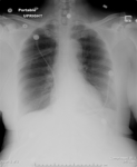
- Title:
- X-ray (chest), PA, Adult Respiratory Distress Syndrome, ARDS, Adult Female
- Description:
- Chest: The endotracheal tube is unchanged in position. There bilateral diffuse pulmonary opacities. There is no pleural effusion. The cardiac silhouette is obscured.
Impression: Progressive diffuse airspace disease compatible with ARDS.
- Keyword:
- X-Ray Radiology, Diagnostic, X-Ray, Diagnostic, Diagnostic X-Ray, ARDS, Human, Diagnosis, Roentgenography, Radiography, Shock Lung, Radiology, Diagnostic X-Ray, Acute Respiratory Distress Syndrome, Diagnostic X-Ray Radiology
- Subject:
- Diagnostic Imaging, Respiratory Tract Diseases, Lung Diseases, Respiratory Distress Syndrome, Adult, Multimodal Imaging, Diagnostic Techniques and Procedures
- Creator:
- Chris DeMauro, MD
- Publisher:
- i-Human Patients, Inc., Inc.
- Language:
- English
- Copyright Holder:
- Chris DeMauro, MD
- Rights:
- http://www.i-human.com/service-agreement-print
- Resource Type:
- Medical Imaging
- Identifier:
- 1328
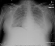
- Title:
- X-ray (chest), AP, Pneumothorax with Chest Tube
- Description:
- Adult female, with chest tube. There is a large right-sided pneumothorax. The left lung is well aerated and clear. The heart is not enlarged. The pulmonary vasculature is not congested.
IMPRESSION: Large right pneumothorax.
- Keyword:
- Primary Spontaneous Pneumothorax, Spontaneous Pneumothorax, Diagnosis, Pressure Pneumothorax, Roentgenography, Tension Pneumothorax, chest tube, Radiology, Diagnostic X-Ray, X-Ray Radiology, Diagnostic, Diagnostic X-Ray Radiology, X-Ray, Diagnostic, Radiography, Diagnostic X-Ray, Pneumothorax
- Subject:
- Diagnostic Techniques and Procedures, Diagnostic Imaging, Respiratory Tract Diseases, Pleural Diseases, Pneumothorax, Multimodal Imaging
- Creator:
- Chris DeMauro, MD
- Publisher:
- i-Human Patients, Inc., Inc.
- Language:
- English
- Copyright Holder:
- Chris DeMauro, MD
- Rights:
- http://www.i-human.com/service-agreement-print
- Resource Type:
- Medical Imaging
- Identifier:
- 1339
- Title:
- X-ray (chest), Lateral, Adult male, Chronic Obstructive Pulmonary Diseaess (COPD)
- Description:
- The lungs are hyperinflated with apical bullous disease. There is flattening of the diaphragm on the lateral view. There is no focal infiltrate. There is no pleural effusion. The heart is mildly enlarged in size. There is no acute bony abnormality.
Impression: Hyperinflation with bullous disease consistent with chronic obstructive pulmonary disease.
- Keyword:
- COPD, Radiography, Chronic Obstructive Pulmonary Disease, Diagnostic X-Ray Radiology, Roentgenography, X-Ray Radiology, Diagnostic, X-Ray, Diagnostic, Chronic Airflow Obstruction, Diagnosis, COAD, Chronic Obstructive Lung Disease, Radiology, Diagnostic X-Ray, Diagnostic X-Ray, Chronic Obstructive Airway Disease
- Subject:
- Pulmonary Disease, Chronic Obstructive, Respiratory Tract Diseases, Lung Diseases, Obstructive, Lung Diseases, Diagnostic Techniques and Procedures, Diagnostic Imaging, Multimodal Imaging
- Creator:
- Chris DeMauro, MD
- Publisher:
- i-Human Patients, Inc., Inc.
- Language:
- English
- Copyright Holder:
- Chris DeMauro, MD
- Rights:
- http://www.i-human.com/service-agreement-print
- Resource Type:
- Medical Imaging
- Identifier:
- 1332
- Title:
- X-ray (chest), PA, Adult Male, Chronic Obstructive Pulmonary Diseaess (COPD)
- Description:
- The lungs are hyperinflated with apical bullous disease. There is flattening of the diaphragm on the lateral view. There is no focal infiltrate. There is no pleural effusion. The heart is mildly enlarged in size. There is no acute bony abnormality.
Impression: Hyperinflation with bullous disease consistent with chronic obstructive pulmonary disease.
- Keyword:
- Chronic Obstructive Airway Disease, X-Ray Radiology, Diagnostic, COAD, COPD, Roentgenography, Chronic Obstructive Lung Disease, X-Ray, Diagnostic, Diagnostic X-Ray, Diagnosis, Chronic Obstructive Pulmonary Disease, Radiology, Diagnostic X-Ray, Radiography, Chronic Airflow Obstruction, Diagnostic X-Ray Radiology
- Subject:
- Diagnostic Techniques and Procedures, Diagnostic Imaging, Multimodal Imaging, Lung Diseases, Obstructive, Pulmonary Disease, Chronic Obstructive, Respiratory Tract Diseases, Lung Diseases
- Creator:
- Chris DeMauro, MD
- Publisher:
- i-Human Patients, Inc., Inc.
- Language:
- English
- Copyright Holder:
- Chris DeMauro, MD
- Rights:
- http://www.i-human.com/service-agreement-print
- Resource Type:
- Medical Imaging
- Identifier:
- 1332
