Search
« Previous |
101 - 110 of 374
|
Next »
Search Results
Select an image to start the slideshow
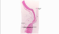
Heart, Semilunar Valve
1 of 10
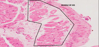
Bundle Of His
2 of 10
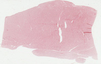
Bundle Of His
3 of 10
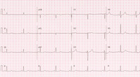
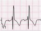
12-Lead ECG: Sinus bradycardia
4 of 10
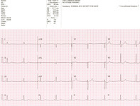
12-Lead ECG: Sinus bradycardia
5 of 10
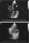
Transthoracic Echocardiogram (TTE), Atrial Septal Defect (ASD)
6 of 10
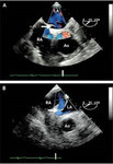
Transthoracic Echocardiogram (TTE), Atrial Septal Defect (ASD)
7 of 10
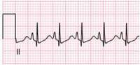
3 Lead ECG: Right Ventricular Hypertrophy (RVH)
8 of 10
3 Lead ECG - Right Ventricular Hypertrophy (RVH)
9 of 10
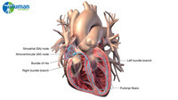
Cardiac Conduction; Labels
10 of 10
