Search
« Previous |
31 - 40 of 301
|
Next »
Search Results
Select an image to start the slideshow
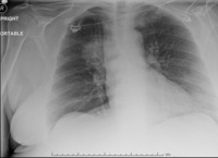
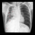
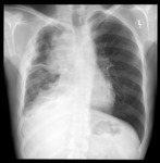
X-ray (chest), AP, Lung Mass Right Upper Lobe (RUL)
1 of 10
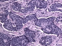
Breast, Invasive Ductal Cancer
2 of 10
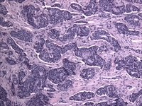
Breast, Invasive Ductal Cancer
3 of 10
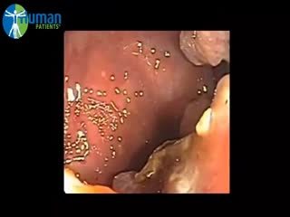
Colonoscopy, Cancer
4 of 10
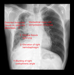
X-ray (chest), PA, Thymoma Invasive, Adult Male
5 of 10
X-ray (chest), PA, Thymoma Invasive, Adult Male
6 of 10

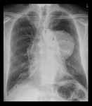
X-ray (chest), PA, Thymoma, Adult Male
7 of 10
X-ray (chest), PA, Thymoma, Adult Male
8 of 10
X-ray (chest), PA, Thymoma, Adult Male
9 of 10
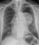
X-ray (chest), PA, Thymoma Invasive, Adult Male
10 of 10
