Search

You searched for: Keyword Pathologic Processes Remove constraint Keyword: Pathologic Processes Language English Remove constraint Language: English

Search Results

  1. Abdominal Pain 3. Epigastric Pain Differential Diagnosis and Approach

    show more
    Description: Epigastric Pain Differential Diagnosis and Approach Duration: 06:55
    Keywords: Diagnosis, Fever, Pathologic Processes, Pathological Conditions. Signs, and Symptoms, Abdominal Pain, Intestine, Large
  2. Abdominal Pain 10. Case Review

    show more
    Description: Abdominal Pain Case Review Duration: 13:35
    Keywords: Intestine, Large, Pathologic Processes, Fever, Diagnosis, Abdominal Pain, Pathological Conditions. Signs, and Symptoms
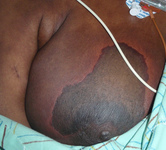
  3. Dermatology: Warfarin-induced Skin Necrosis, Fatal, Breast

    show more
    Description: Fatal skin necrosis at breast, induced by heparin. Skin necrosis is the pathologic death of one or more cells, or of a portion of tissue or organ, resulting from irreversible damage; earliest irreversible changesare mitochondrial, consisting of swelling and granular calcium deposits seen by electron microscopy; most frequent visible alterationsare nuclear: pyknosis, shrunken and abnormally dark basophilic staining; karyolysis, swollen and abnormally pale basophilic staining;or karyorrhexis, rupture, and fragmentation of the nucleus. After such changes, the outlines of individual cells are indistinct, andaffected cells may merge, sometimes forming a focus of coarsely granular, amorphous, or hyaline material.
    Keywords: skin, Cell death, Pathological Conditions, mitochondrial swelling, cell lysis, Pathologic Processes, Signs and Symptoms
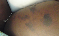
  4. Dermatology: Skin, Warfarin-induced Skin Necrosis, Fatal

    show more
    Description: Fatal skin necrosis, induced by heparin. Skin necrosis is the pathologic death of one or more cells, or of a portion of tissue or organ, resulting from irreversible damage; earliest irreversible changesare mitochondrial, consisting of swelling and granular calcium deposits seen by electron microscopy; most frequent visible alterationsare nuclear: pyknosis, shrunken and abnormally dark basophilic staining; karyolysis, swollen and abnormally pale basophilic staining;or karyorrhexis, rupture, and fragmentation of the nucleus. After such changes, the outlines of individual cells are indistinct, andaffected cells may merge, sometimes forming a focus of coarsely granular, amorphous, or hyaline material
    Keywords: mitochondrial swelling, Signs and Symptoms, Pathological Conditions, skin, Cell death, Pathologic Processes, cell lysis
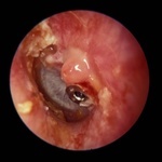
  5. Tube Granuloma, Tympanic Membrane, Image 4

    show more
    Description: Tympanic membrane - Tube granuloma 4 By definition, a granuloma is a nodular inflammatory lesion. They are usually small and consist primarily of compact mononuclear phagocytes. Granulomas are different from other inflammatory reactions; they represent an ultimately unsuccessful localized attempt to rid the host of an offending organism or process. Granulomatous diseases in the middle ear may be localized primarily to the ear and surrounding tissues, or they may be a manifestation of a body-wide disseminated problem.
    Keywords: Ear, Signs and Symptoms, mononuclear phagocytes, nodular inflammatory lesion, Pathological Conditions, Pathologic Processes
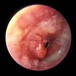
  6. Tube Granuloma, Tympanic Membrane, Image 3

    show more
    Description: Tympanic membrane - Tube granuloma 3 By definition, a granuloma is a nodular inflammatory lesion. They are usually small and consist primarily of compact mononuclear phagocytes. Granulomas are different from other inflammatory reactions; they represent an ultimately unsuccessful localized attempt to rid the host of an offending organism or process. Granulomatous diseases in the middle ear may be localized primarily to the ear and surrounding tissues, or they may be a manifestation of a body-wide disseminated problem.
    Keywords: Signs and Symptoms, mononuclear phagocytes, Pathological Conditions, Pathologic Processes, Ear, nodular inflammatory lesion
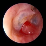
  7. Tube Granuloma, Tympanic Membrane, Image 1

    show more
    Description: Tympanic membrane - Tube granuloma 1 By definition, a granuloma is a nodular inflammatory lesion. They are usually small and consist primarily of compact mononuclear phagocytes. Granulomas are different from other inflammatory reactions; they represent an ultimately unsuccessful localized attempt to rid the host of an offending organism or process. Granulomatous diseases in the middle ear may be localized primarily to the ear and surrounding tissues, or they may be a manifestation of a body-wide disseminated problem.
    Keywords: nodular inflammatory lesion, Pathologic Processes, Signs and Symptoms, Ear, Pathological Conditions, mononuclear phagocytes
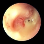
  8. Tube Granuloma, Tympanic Membrane, Image 2

    show more
    Description: Tympanic membrane - Tube granuloma 2 By definition, a granuloma is a nodular inflammatory lesion. They are usually small and consist primarily of compact mononuclear phagocytes. Granulomas are different from other inflammatory reactions; they represent an ultimately unsuccessful localized attempt to rid the host of an offending organism or process. Granulomatous diseases in the middle ear may be localized primarily to the ear and surrounding tissues, or they may be a manifestation of a body-wide disseminated problem.
    Keywords: Signs and Symptoms, Ear, Pathological Conditions, nodular inflammatory lesion, mononuclear phagocytes, Pathologic Processes