Search
« Previous |
11 - 20 of 34
|
Next »
Search Results
Select an image to start the slideshow
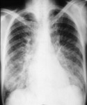
X-ray (chest), PA, Pulmonary Edema Lower Lobes, Adult Male
1 of 10
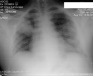
X-ray (chest), PA, Hospital Acquired Pneumonia, Adult Female
2 of 10
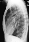
X-ray (chest), PA and Lateral, Chronic Obstructive Pulmonary Disease (COPD), Adult Female
3 of 10
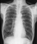
X-ray (chest), PA, Chronic Obstructive Pulmonary Disease (COPD), Adult Female
4 of 10

X-ray (chest), AP, Bilateral Mid-and-Lower Lobe Infiltrates
5 of 10
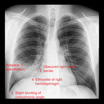
X-ray (chest), PA, Pneumonia RML Silhouette Sign, Answers, Adult Male
6 of 10
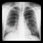
X-ray (chest), PA, Pneumonia RML Silhouette Sign, Numbered, Adult Male
7 of 10
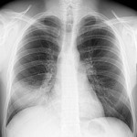
X-ray (chest), PA, Pneumonia RML Silhouette Sign, Adult Male
8 of 10
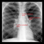
X-ray (chest), PA, Calcified Mediastinal Nodes, Adult Male
9 of 10

X-ray (chest), PA, Calcified Mediastinal Nodes, Adult Male
10 of 10
