Search
« Previous |
21 - 30 of 51
|
Next »
Search Results

- Title:
- X-ray (chest), Cavitary Lesion of the Lung in Cystic Fibrosis
- Description:
- Chest, frontal and lateral, upright, 43 yo F, ALL
- Keyword:
- Diagnostic X-Ray, Pulmonary Cystic Fibrosis, X-Ray, Diagnostic, Diagnosis, Cavitary Lesion of the Lung/Cystic Fibrosis, X-Ray Radiology, Diagnostic, Roentgenography, Radiography, Diagnostic X-Ray Radiology, Radiology, Diagnostic X-Ray
- Subject:
- Diagnostic Imaging, Diagnostic Techniques and Procedures, Multimodal Imaging, Lung Diseases, Respiratory Tract Diseases, Cystic Fibrosis
- Creator:
- Rush University Medical Center
- Contributor:
- i-Human-Rush radiology project interns
- Publisher:
- Rush University Medical Center
- Language:
- English
- Copyright Holder:
- Rush Medical College
- Rights:
- http://www.i-human.com/service-agreement-print
- Resource Type:
- Medical Imaging

- Title:
- X-ray (chest), Cavitary Lesion of the Lung in Cystic Fibrosis
- Description:
- Chest, frontal and lateral, upright, 43 yo F, ALL
- Keyword:
- Radiography, X-Ray, Diagnostic, X-Ray Radiology, Diagnostic, Cavitary Lesion of the Lung/Cystic Fibrosis, Diagnostic X-Ray Radiology, Diagnostic X-Ray, Radiology, Diagnostic X-Ray, Pulmonary Cystic Fibrosis, Diagnosis, Roentgenography
- Subject:
- Lung Diseases, Diagnostic Imaging, Cystic Fibrosis, Respiratory Tract Diseases, Diagnostic Techniques and Procedures, Multimodal Imaging
- Creator:
- Rush University Medical Center
- Contributor:
- i-Human-Rush radiology project interns
- Publisher:
- Rush University Medical Center
- Language:
- English
- Copyright Holder:
- Rush Medical College
- Rights:
- http://www.i-human.com/service-agreement-print

- Title:
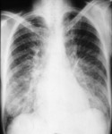
- X-ray (chest), PA, Pneumonia, Left Lower Lobe
- Description:
- CXR PA - Pneumonia left lower lobe
- Keyword:
- Lung Inflammation, Pulmonary Inflammation, Pneumonia, Lobar, Pneumonitis, Diagnostic X-Ray Radiology, Experimental Lung Inflammation, Diagnostic X-Ray, Radiology, Diagnostic X-Ray, X-Ray, Diagnostic, Radiography, Diagnosis, X-Ray Radiology, Diagnostic, Roentgenography, Lobar Pneumonia, Thoracic Radiography
- Subject:
- Pneumonia, Lung Diseases, Diagnostic Techniques and Procedures, Respiratory Tract Diseases, Diagnostic Imaging, Multimodal Imaging, Radiography, Thoracic
- Creator:
- James Carlson, PhD, PACRosalind Franklin University
- Publisher:
- Rosalind Franklin University School of Medicine
- Language:
- English
- Copyright Holder:
- Rosalind Franklin University School of Medicine
- Rights:
- http://www.i-human.com/service-agreement-print
- Resource Type:
- Medical Imaging
- Identifier:
- 1782
- Title:
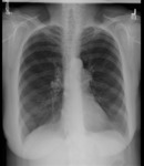
- X-ray (chest), PA and Lateral, Chronic Obstructive Pulmonary Disease (COPD), Adult Female
- Description:
- Posterior-anterior and Lateral views of the chest reveal clear lung fields, normal cardiac silhouette, but with hyperexpansion and flattened diaphragms consistent with obstructive airways disease such as emphysema or asthma. No infiltrate, mass, or pulmonary vascular congestion is seen.
- Keyword:
- X-Ray, Diagnostic, Radiology, Diagnostic X-Ray, Chronic Obstructive Airway Disease, Radiography, Diagnostic X-Ray Radiology, Diagnostic X-Ray, COPD, Roentgenography, Diagnosis, X-Ray Radiology, Diagnostic, Chronic Airflow Obstruction, Chronic Obstructive Lung Disease, Chronic Obstructive Pulmonary Disease, COAD
- Subject:
- Diagnostic Techniques and Procedures, Respiratory Tract Diseases, Lung Diseases, Lung Diseases, Obstructive, Multimodal Imaging, Pulmonary Disease, Chronic Obstructive, Diagnostic Imaging
- Creator:
- Jah-Won Koo, MDRush Medical College
- Publisher:
- Rush Medical College
- Language:
- English
- Copyright Holder:
- Rush Medical College
- Rights:
- http://www.i-human.com/service-agreement-print
- Resource Type:
- Medical Imaging
- Identifier:
- 1718
- Title:
- X-ray (chest), PA and Lateral, Chronic Obstructive Pulmonary Disease (COPD), Adult Female
- Description:
- Posterior-anterior and Lateral views of the chest reveal clear lung fields, normal cardiac silhouette, but with hyperexpansion and flattened diaphragms consistent with obstructive airways disease such as emphysema or asthma. No infiltrate, mass, or pulmonary vascular congestion is seen.
- Keyword:
- Roentgenography, Diagnosis, Chronic Obstructive Lung Disease, Diagnostic X-Ray Radiology, Diagnostic X-Ray, Chronic Obstructive Pulmonary Disease, Radiology, Diagnostic X-Ray, Radiography, COAD, Chronic Airflow Obstruction, X-Ray, Diagnostic, COPD, Chronic Obstructive Airway Disease, X-Ray Radiology, Diagnostic
- Subject:
- Respiratory Tract Diseases, Diagnostic Techniques and Procedures, Lung Diseases, Obstructive, Multimodal Imaging, Pulmonary Disease, Chronic Obstructive, Lung Diseases, Diagnostic Imaging
- Creator:
- Jah-Won Koo, MDRush Medical College
- Publisher:
- Rush Medical College
- Language:
- English
- Copyright Holder:
- Rush Medical College
- Rights:
- http://www.i-human.com/service-agreement-print
- Resource Type:
- Medical Imaging
- Identifier:
- 1718
- Title:
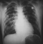
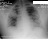
- X-ray (chest), AP, Pulmonary Infiltrate, Mitral Insufficiency / Regurgitation
- Description:
- The chest x-ray of an adult male shows a normal cardiac silhouette with a diffuse infiltrate throughout both lung fields suggesting pulmonary congestion secondary to mitral valve regurgitation.
- Keyword:
- pulmonary congestion, Diagnostic X-Ray Radiology, Roentgenography, Diagnosis, Radiography, Radiology, Diagnostic X-Ray, X-Ray, Diagnostic, pumonary infiltrate, X-Ray Radiology, Diagnostic, Diagnostic X-Ray
- Subject:
- Lung Diseases, Multimodal Imaging, Diagnostic Imaging, Respiratory Tract Diseases, Pulmonary Edema, Diagnostic Techniques and Procedures
- Creator:
- Avery Ellis, M.D., Ph.D.University of Buffalo School of Medicine
- Publisher:
- University of Buffalo School of Medicine
- Language:
- English
- Copyright Holder:
- University at Buffalo School of Medicine
- Rights:
- http://www.i-human.com/service-agreement-print
- Identifier:
- 1697
- Title:
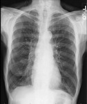
- X-ray (chest), PA, Chronic Obstructive Pulmonary Disease (COPD), Adult Male
- Description:
- Hyperinflation, flattened diaphragm, no infiltrates or effusions
- Keyword:
- Chronic Airflow Obstruction, Diagnostic X-Ray Radiology, X-Ray Radiology, Diagnostic, COPD, Chronic Obstructive Lung Disease, Diagnosis, Radiology, Diagnostic X-Ray, Roentgenography, Radiography, COAD, Diagnostic X-Ray, Chronic Obstructive Airway Disease, Chronic Obstructive Pulmonary Disease, X-Ray, Diagnostic
- Subject:
- Diagnostic Imaging, Multimodal Imaging, Lung Diseases, Obstructive, Diagnostic Techniques and Procedures, Pulmonary Disease, Chronic Obstructive, Lung Diseases, Respiratory Tract Diseases
- Creator:
- Susan Gallagher, MD University of Buffalo School of Medicine
- Publisher:
- i-Human Patients, Inc.
- Language:
- English
- Copyright Holder:
- Susan Gallagher, MD
- Rights:
- http://www.i-human.com/service-agreement-print
- Resource Type:
- Medical Imaging
- Identifier:
- 1695
- Title:
- X-ray (chest), PA, Pulmonary Edema Lower Lobes, Adult Male
- Description:
- CXR PA - Adult Male, pulmonary edema lower lobes
- Keyword:
- X-Ray, Diagnostic, Radiography, Wet Lung, Diagnostic X-Ray, Radiology, Diagnostic X-Ray, X-Ray Radiology, Diagnostic, Pulmonary Edemas, Diagnostic X-Ray Radiology, Diagnosis, Edemas, Pulmonary, Edema, Pulmonary, Roentgenography
- Subject:
- Respiratory Tract Diseases, Diagnostic Imaging, Pulmonary Edema, Multimodal Imaging, Lung Diseases, Diagnostic Techniques and Procedures
- Creator:
- Jah-Won KooRush Medical College
- Publisher:
- Rush Medical College
- Language:
- English
- Copyright Holder:
- Rush Medical College
- Rights:
- http://www.i-human.com/service-agreement-print
- Resource Type:
- Medical Imaging
- Identifier:
- 1687
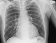
- Title:
- X-ray (chest), PA, Hospital Acquired Pneumonia, Adult Female
- Description:
- Partial consolidation of the right upper lobe; scattered left lung patchy opacities.
- Keyword:
- Radiography, Radiology, Diagnostic X-Ray, Diagnostic X-Ray, Pneumonitis, Pneumonia, Lobar, X-Ray, Diagnostic, Pulmonary Inflammation, Lobar Pneumonia, Lung Inflammation, Experimental Lung Inflammation, Roentgenography, Diagnostic X-Ray Radiology, X-Ray Radiology, Diagnostic, Diagnosis
- Subject:
- Diagnostic Techniques and Procedures, Pneumonia, Respiratory Tract Diseases, Lung Diseases, Multimodal Imaging, Diagnostic Imaging
- Creator:
- Jennifer Babik, MD, PhDUCSF Dept of Medicine
- Publisher:
- UCSF Dept of Medicine
- Language:
- English
- Copyright Holder:
- UCSF
- Rights:
- http://www.i-human.com/service-agreement-print
- Resource Type:
- Medical Imaging
- Identifier:
- 1658
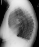
- Title:
- X-ray (chest), PA and Lateral, Chronic Obstructive Pulmonary Disease (COPD), Adult Female
- Description:
- Increased AP diameter, flattening of the diaphragm, decreased lung markings
- Keyword:
- COAD, Diagnosis, Pulmonary Disease, Chronic Obstructive, Chronic Obstructive Pulmonary Disease, Diagnostic X-Ray Radiology, Diagnostic X-Ray, X-Ray Radiology, Diagnostic, Radiology, Diagnostic X-Ray, Chronic Obstructive Airway Disease, Roentgenography, Chronic Airflow Obstruction, X-Ray, Diagnostic, Chronic Obstructive Lung Disease, COPD, Radiography
- Subject:
- Lung Diseases, Obstructive, Lung Diseases, Pulmonary Disease, Chronic Obstructive, Diagnostic Techniques and Procedures, Multimodal Imaging, Respiratory Tract Diseases, Diagnostic Imaging
- Creator:
- James Heilman, MD
- Publisher:
- Wikimedia Commons https://commons.wikimedia.org/wiki/File:COPD.JPG
- Language:
- English
- Rights:
- https://creativecommons.org/licenses/by-sa/3.0
- Resource Type:
- Medical Imaging
- Identifier:
- 1656
