Search
Search Results
Select an image to start the slideshow
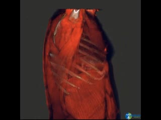
CT (chest), Thoracic Anatomy Reconstruction 3D
1 of 6
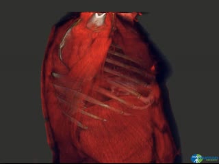
CT Volume Reconstruction Heart, Lungs, Thoracic Muscles, Rib Cage
2 of 6
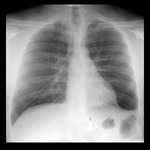
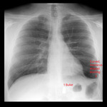
X-ray (chest), AP, Adult Male, Thoracic Bullet, Annotated
3 of 6
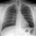
X-ray (chest), LAT, Adult Male, Thoracic Bullet
4 of 6
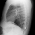
X-ray (chest), LAT, Adult Male, Thoracic Bullet
5 of 6
X-ray (chest), AP, Adult Male, Thoracic Bullet, Annotated Answers
6 of 6
