×
You need to sign in or sign up before continuing.
Search
« Previous |
61 - 70 of 127
|
Next »
Search Results
- Title:
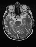
- MRI Brain - Alzheimer's Disease
- Description:
- Findings:
Pronounced brain volume decrease, mostly supratentorial with significant reduction of hippocampal volume, and increased signal in T2 sequence. This increase is clear for hippocampal head, body and tail in flair sequences.
Small gliosis foci in white matter of semioval centers.
No signs of hemorrhage or dural collections.
Gadolinium contrast doesnt show any other pathology
Conclusion: Significant bilateral hippocampal atrophy, suggestive of Alzheimers disease.
- Keyword:
- cytopathology, Pons, pathology, Multiple Sclerosis, Dementia, Acute Confusional Senile Dementia, Demyelinating Autoimmune Disease, Autoimmune, Delirium, Amnestic, histopathology, Cognitive Disorders, Brain, Alzheimer Disease, biopsy
- Subject:
- Neurodegenerative Diseases, Central Nervous System Diseases, Central Nervous System, Nervous System Diseases, Staining and Labeling, Histological Techniques, Alzheimer Disease, Nervous System, Brain, Mental Disorders, Histocytological Preparation Techniques
- Creator:
- Jaime Drewes MD
- Publisher:
- i-Human Patients, Inc.
- Language:
- English
- Copyright Holder:
- Jaime Drewes MD
- Rights:
- http://www.i-human.com/service-agreement-print
- Resource Type:
- Medical Imaging
- Identifier:
- 3166
- Title:
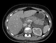
- CT (abdomen), Acute Pancreatitis
- Description:
- The CT shows pancreas of increased volume, and heterogeneous structure with blurred contour. Abundant exudate can be found surrounding it.
- Keyword:
- Acute Necrotizing Pancreatitis, Pancreatitis, Acute Necrotizing, Necrotizing Pancreatitis, Acute, digestive system, histopathology, pancreatitis, Acute Pain, endocrine gland, necrotizing hemorrhagic pancreatitis, pancreas, Pathology, biopsy, cytopathology
- Subject:
- Pancreatitis, Staining and Labeling, Histocytological Preparation Techniques, Digestive System Diseases, Pancreatitis, Acute Necrotizing, Pancreatic Diseases, Histological Techniques, Hemorrhage
- Creator:
- Jaime Drewes MD
- Publisher:
- Jaime Drewes MD
- Language:
- English
- Rights:
- http://www.i-human.com/service-agreement-print
- Resource Type:
- Medical Imaging
- Identifier:
- 3164
- Title:
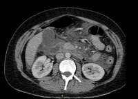
- CT (abdomen), Acute Pancreatitis
- Description:
- The CT shows pancreas of increased volume, and heterogeneous structure with blurred contour. Abundant exudate can be found surrounding it.
- Keyword:
- necrotizing hemorrhagic pancreatitis, biopsy, cytopathology, pancreas, Pancreatitis, Acute Necrotizing, digestive system, histopathology, endocrine gland, Pathology, Acute Pain, Necrotizing Pancreatitis, Acute, Acute Necrotizing Pancreatitis, pancreatitis
- Subject:
- Histocytological Preparation Techniques, Pancreatic Diseases, Digestive System Diseases, Histological Techniques, Staining and Labeling, Pancreatitis, Acute Necrotizing, Pancreatitis, Hemorrhage
- Creator:
- Jaime Drewes MD
- Publisher:
- Jaime Drewes MD
- Language:
- English
- Rights:
- http://www.i-human.com/service-agreement-print
- Resource Type:
- Medical Imaging
- Identifier:
- 3164
- Title:
- CT (abdomen), Acute Pancreatitis
- Description:
- The CT shows pancreas of increased volume, and heterogeneous structure with blurred contour. Abundant exudate can be found surrounding it.
- Keyword:
- necrotizing hemorrhagic pancreatitis, Acute Pain, Acute Necrotizing Pancreatitis, endocrine gland, pancreatitis, pancreas, Pancreatitis, Acute Necrotizing, cytopathology, Necrotizing Pancreatitis, Acute, biopsy, digestive system, histopathology, Pathology
- Subject:
- Hemorrhage, Pancreatitis, Digestive System Diseases, Histocytological Preparation Techniques, Pancreatitis, Acute Necrotizing, Staining and Labeling, Pancreatic Diseases, Histological Techniques
- Creator:
- Jaime Drewes MD
- Publisher:
- Jaime Drewes MD
- Language:
- English
- Rights:
- http://www.i-human.com/service-agreement-print
- Resource Type:
- Medical Imaging
- Identifier:
- 3164
- Title:
- Malaria, blood
- Description:
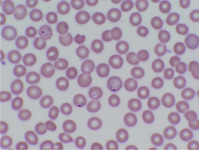
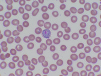
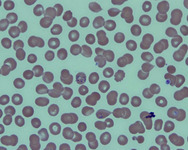
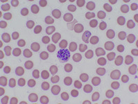
- Malaria, blood
Malaria 1: Ring forms of P vivax. An infected RBC in the center of the smear also shows Schuffners dots.
Malaria 2: P. vivax gametocyte.
Malaria 3: P vivax schizont showing >12 merozoites.
Malaria 4: Ring forms of P. vivax.
- Keyword:
- pathology, Parasitic Diseases, Malaria, Viral Diseases, histopathology, cytopathology, Protozoan Infections, biopsy, Protozoan
- Subject:
- Histological Techniques, Staining and Labeling, Parasitic Diseases, Histocytological Preparation Techniques, Malaria
- Creator:
- Jennifer Babik, MD
- Publisher:
- Jenifer Babik, MD
- Language:
- English
- Rights:
- http://www.i-human.com/service-agreement-print
- Resource Type:
- Slide
- Identifier:
- 3116
- Title:
- Blood, malaria
- Description:
- Malaria, blood
Malaria 1: Ring forms of P vivax. An infected RBC in the center of the smear also shows Schuffners dots.
Malaria 2: P. vivax gametocyte.
Malaria 3: P vivax schizont showing >12 merozoites.
Malaria 4: Ring forms of P. vivax.
- Keyword:
- Protozoan Infections, Viral Diseases, pathology, Malaria, Parasitic Diseases, cytopathology, biopsy, Protozoan, histopathology
- Subject:
- Parasitic Diseases, Staining and Labeling, Malaria, Histological Techniques, Histocytological Preparation Techniques
- Creator:
- Jennifer Babik, MD
- Publisher:
- Jenifer Babik, MD
- Language:
- English
- Rights:
- http://www.i-human.com/service-agreement-print
- Resource Type:
- Slide
- Identifier:
- 3116
- Title:
- Blood, malaria
- Description:
- Malaria, blood
Malaria 1: Ring forms of P vivax. An infected RBC in the center of the smear also shows Schuffners dots.
Malaria 2: P. vivax gametocyte.
Malaria 3: P vivax schizont showing >12 merozoites.
Malaria 4: Ring forms of P. vivax.
- Keyword:
- Malaria, cytopathology, Parasitic Diseases, histopathology, Protozoan, biopsy, pathology, Viral Diseases, Protozoan Infections
- Subject:
- Histological Techniques, Parasitic Diseases, Staining and Labeling, Malaria, Histocytological Preparation Techniques
- Creator:
- Jennifer Babik, MD
- Publisher:
- Jenifer Babik, MD
- Language:
- English
- Rights:
- http://www.i-human.com/service-agreement-print
- Resource Type:
- Slide
- Identifier:
- 3116
- Title:
- Malaria, blood
- Description:
- Malaria, blood
Malaria 1: Ring forms of P vivax. An infected RBC in the center of the smear also shows Schuffners dots.
Malaria 2: P. vivax gametocyte.
Malaria 3: P vivax schizont showing >12 merozoites.
Malaria 4: Ring forms of P. vivax.
- Keyword:
- cytopathology, Parasitic Diseases, Malaria, Viral Diseases, pathology, Protozoan, Protozoan Infections, biopsy, histopathology
- Subject:
- Histological Techniques, Parasitic Diseases, Histocytological Preparation Techniques, Malaria, Staining and Labeling
- Creator:
- Jennifer Babik, MD
- Publisher:
- Jenifer Babik, MD
- Language:
- English
- Rights:
- http://www.i-human.com/service-agreement-print
- Resource Type:
- Slide
- Identifier:
- 3116
- Title:
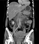
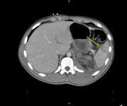
- CT (abdomen), Splenic Rupture
- Description:
- CT Scan showing traumatic splenic rupture on its anterior superior area. Green arrow highlights the fracture
- Keyword:
- cytopathology, biopsy, splenic rupture, pathology, histopathology, rupture, spleen
- Subject:
- Histocytological Preparation Techniques, Splenic Diseases, Splenic Rupture, Staining and Labeling, Histological Techniques, Lymphatic Diseases, Hemic and Lymphatic Diseases
- Creator:
- Jaime Drewes MD
- Publisher:
- Jaime Drewes MD
- Language:
- English
- Rights:
- http://www.i-human.com/service-agreement-print
- Resource Type:
- Medical Imaging
- Identifier:
- 3080

- Title:
- CT (abdomen), Splenic Rupture
- Description:
- CT Scan showing traumatic splenic rupture on its anterior superior area. Green arrow highlights the fracture
- Keyword:
- cytopathology, histopathology, rupture, biopsy, spleen, pathology, splenic rupture
- Subject:
- Splenic Rupture, Histological Techniques, Histocytological Preparation Techniques, Lymphatic Diseases, Splenic Diseases, Staining and Labeling, Hemic and Lymphatic Diseases
- Creator:
- Jaime Drewes MD
- Publisher:
- Jaime Drewes MD
- Language:
- English
- Rights:
- http://www.i-human.com/service-agreement-print
- Resource Type:
- Medical Imaging
- Identifier:
- 3080
