Search
« Previous |
81 - 90 of 94
|
Next »
Search Results
Select an image to start the slideshow

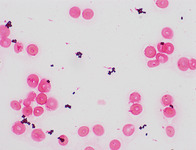
Blood, Peripheral Blood Smear
1 of 10
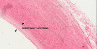
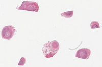
Subintima Thickening
2 of 10
Subintima Thickening
3 of 10

Subintima Thickening
4 of 10
Throat, Swab Group A Streptococcus
5 of 10
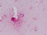
Staphylococcus Aureus Blood Culture, Gram Stain
6 of 10
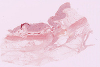
Temporal Artery, Giant Cell Arteritis
7 of 10

Bone Marrow, Multiple Myeloma
8 of 10

Blood, Thrombotic Thrombocytic Purpura
9 of 10
Stomach, Peptic Ulcer
10 of 10
