Search
Search Results
- Title:
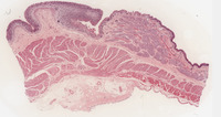
- Gastro-esophageal Junction
- Description:
- System: Gastrointestinal
Organ: Esophageal cardiac junction
Disease process: Normal
Species: Human
Highest magnification: 20x
Stain: H&E
Note the epithelial transition from stratified squamous non-keratinized (esophagus) to simple columnar (gastric). There are two sets of glands: esophageal glands (in the submucosa) vs. cardiac gastric glands (in the mucosa).
In the muscularis layer the “sphincter” is an expansion of the middle oblique layer in the gastric muscularis which forms an incomplete sphincter, i.e., “sling” or valve + skeletal muscle (crura of diaphragm).
The cells that line the gastric glands in the cardiac region of the stomach: they are all mucous producing cells. The luminal surface and the gastric pits are also lined by surface mucus cells.
- Keyword:
- histology, small intestine, Anatomy & histology, digestive system, gastro-esophageal junction, morphology, anatomy, esophageal-cardiac junction
- Subject:
- Histocytological Preparation Techniques, Histological Techniques, Staining and Labeling, Esophagogastric Junction
- Creator:
- Rush Medical College
- Publisher:
- Rush Medical College
- Language:
- English
- Copyright Holder:
- Rush Medical College
- Rights:
- http://www.i-human.com/service-agreement-print
- Resource Type:
- Slide
- Identifier:
- 168
