Search
Search Results
Select an image to start the slideshow
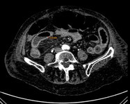
CT Abdomen (axial), Acute Mesenteric Ischemia
1 of 9
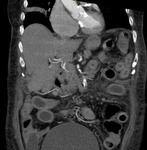
CT Abdomen - Acute Mesenteric Ischemia
2 of 9
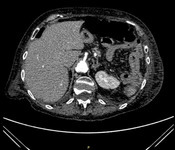
CT (abdomen), Acute Mesenteric Ischemia
3 of 9
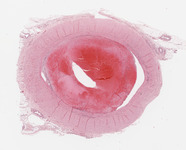
Vein, Organizing and recanalizing thrombus
4 of 9
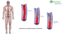
Vascular Embolus/thrombus
5 of 9

Embolus Or Thrombus Mesenteric Ischemia
6 of 9
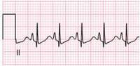
3 Lead ECG: Right Ventricular Hypertrophy (RVH)
7 of 9
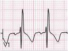
3 Lead ECG - Right Ventricular Hypertrophy (RVH)
8 of 9
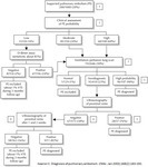
Diagnostic algorithm: Pulmonary embolism
9 of 9
