Search
« Previous |
11 - 20 of 93
|
Next »
Search Results
Select an image to start the slideshow
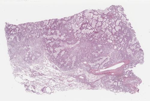
Lung, Bronchial carcinoma, adenocarcinoma
1 of 10
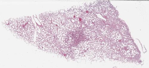
Lung, Interstitial pulmonary fibrosis
2 of 10
Dyspnea - Chronic pulmonary embolism
3 of 10

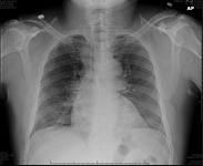
Chest Pain - Pulmonary Embolism
4 of 10
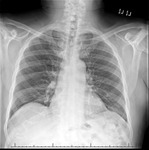
X-ray (chest), PA, Adult Male, Normal
5 of 10
X-ray (chest), AP, Adult Male, Normal
6 of 10
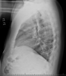
X-ray (chest), Lateral, Adult Male, Normal
7 of 10
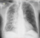
CXR AP-L Interstitial Lung Disease
8 of 10
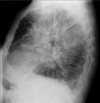
CXR AP-L Interstitial Lung Disease
9 of 10

CT (chest), Interstitial Lung Disease (ILD)
10 of 10
