×
You need to sign in or sign up before continuing.
Search
« Previous |
31 - 40 of 87
|
Next »
Search Results
- Title:
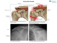
- Shoulder Trauma
- Description:
- 2 x 2 illustration and radiograph set showing a normal shoulder vs. various examples of shoulder trauma, including clavicular fracture, humeral head fracture at the surgical neck, and rotator cuff tear
- Keyword:
- clavicle, shoulder trauma, shoulder, Fractures, Bone, surgical neck of the humerus, rotator cuff, clavicular fracture
- Subject:
- Wounds and Injuries, Clavicle, Humerus
- Creator:
- Laura Garrison
i-Human Patients, Inc.
- Publisher:
- i-Human Patients, Inc.
- Language:
- English
- Copyright Holder:
- i-Human Patients, Inc.
- Rights:
- All rights reserved
- Resource Type:
- Illustration
- Identifier:
- 2376
- Title:
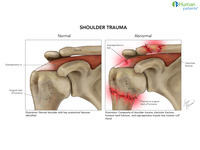
- Shoulder Trauma
- Description:
- Illustration set showing a normal shoulder vs. various examples of shoulder trauma, including clavicular fracture, humeral head fracture at the surgical neck, and rotator cuff tear
- Keyword:
- Fractures, Bone, rotator cuff, clavicular fracture, shoulder trama, shoulder
- Subject:
- Wounds and Injuries, Humerus, Clavicle
- Creator:
- Laura Garrisoni-Human Patients, Inc.
- Publisher:
- i-Human Patients, Inc.
- Language:
- English
- Copyright Holder:
- i-Human Patients, Inc.
- Rights:
- http://www.i-human.com/service-agreement-print
- Resource Type:
- Illustration
- Identifier:
- 2377
- Title:
- X-ray (foot), Image 2, Lisfranc Fracture Dislocation
- Description:
- X-ray right foot 3 views complete, 33 yo M, all
- Keyword:
- X-Ray, Diagnostic, tarsometatarsal injury, midfoot injury, Diagnostic X-Ray, Lisfranc fracture, X-Ray Radiology, Diagnostic, Lisfranc dislocation, Radiography, Diagnostic X-Ray Radiology, Diagnosis, Radiology, Diagnostic X-Ray, Lisfranc Fracture Dislocation, Lisfranc injury, Roentgenography
- Subject:
- Extremities, Multimodal Imaging, Wounds and Injuries, Body Regions, Diagnostic Imaging, Dislocations, Foot, Lower Extremity, Diagnostic Techniques and Procedures
- Creator:
- Rush University Medical Center
- Contributor:
- i-Human-Rush radiology project interns
- Publisher:
- Rush University Medical Center
- Language:
- English
- Copyright Holder:
- Rush Medical College
- Rights:
- http://www.i-human.com/service-agreement-print
- Resource Type:
- Medical Imaging
- Title:
- X-ray (foot), Image 1, Lisfranc Fracture Dislocation
- Description:
- X-ray right foot 3 views complete, 33 yo M, all
- Keyword:
- Lisfranc dislocation, tarsometatarsal injury, Radiography, X-Ray Radiology, Diagnostic, midfoot injury, Diagnostic X-Ray, Diagnostic X-Ray Radiology, Lisfranc injury, Lisfranc Fracture Dislocation, Roentgenography, X-Ray, Diagnostic, Lisfranc fracture, Radiology, Diagnostic X-Ray, Diagnosis
- Subject:
- Extremities, Body Regions, Lower Extremity, Multimodal Imaging, Wounds and Injuries, Foot, Dislocations, Diagnostic Imaging, Diagnostic Techniques and Procedures
- Creator:
- Rush University Medical Center
- Contributor:
- i-Human-Rush radiology project interns
- Publisher:
- Rush University Medical Center
- Language:
- English
- Copyright Holder:
- Rush Medical College
- Rights:
- http://www.i-human.com/service-agreement-print
- Resource Type:
- Medical Imaging

- Title:
- X-ray (plevis), Pelvic Fracture, Male
- Description:
- Xray - pelvic fracture, male
- Keyword:
- Diagnosis, Radiography, Roentgenography, Diagnostic X-Ray Radiology, X-Ray Radiology, Diagnostic, Diagnostic X-Ray, Radiology, Diagnostic X-Ray, X-Ray, Diagnostic
- Subject:
- Fractures, Bone, Diagnostic Imaging, Multimodal Imaging, Wounds and Injuries, Diagnostic Techniques and Procedures
- Creator:
- i-Human Patients, Inc
- Publisher:
- i-Human Patients, Inc.
- Language:
- English
- Copyright Holder:
- i-Human Patients, Inc.
- Rights:
- http://www.i-human.com/service-agreement-print
- Resource Type:
- Medical Imaging
- Identifier:
- 2235
- Title:
- X-ray (knee), Image 3, Comminuted Fracture of Patella
- Description:
- X-ray Left knee complete, 71 yo F, all
- Keyword:
- knee fracture, Comminuted fracture of patella, Radiography, Diagnostic X-Ray, kneecap, X-Ray, Diagnostic, Diagnostic X-Ray Radiology, Roentgenography, Diagnosis, Radiology, Diagnostic X-Ray, X-Ray Radiology, Diagnostic
- Subject:
- Multimodal Imaging, Wounds and Injuries, Diagnostic Techniques and Procedures, Diagnostic Imaging, Patella, Fractures, Bone
- Creator:
- Rush University Medical Center
- Contributor:
- i-Human-Rush radiology project interns
- Publisher:
- Rush University Medical Center
- Language:
- English
- Copyright Holder:
- Rush Medical College
- Rights:
- http://www.i-human.com/service-agreement-print
- Resource Type:
- Medical Imaging

- Title:
- X-ray (knee), Image 2, Comminuted Fracture of Patella
- Description:
- X-ray Left knee complete, 71 yo F, all
- Keyword:
- Comminuted fracture of patella, X-Ray, Diagnostic, Radiography, knee fracture, Radiology, Diagnostic X-Ray, X-Ray Radiology, Diagnostic, Roentgenography, Diagnostic X-Ray Radiology, kneecap, Diagnosis, Diagnostic X-Ray
- Subject:
- Wounds and Injuries, Fractures, Bone, Patella, Diagnostic Imaging, Diagnostic Techniques and Procedures, Multimodal Imaging
- Creator:
- Rush University Medical Center
- Contributor:
- i-Human-Rush radiology project interns
- Publisher:
- Rush University Medical Center
- Language:
- English
- Copyright Holder:
- Rush Medical College
- Rights:
- http://www.i-human.com/service-agreement-print
- Resource Type:
- Medical Imaging
- Title:
- X-ray (knee), Image 1, Comminuted Fracture of Patella
- Description:
- X-ray Left knee complete, 71 yo F, all
- Keyword:
- X-Ray Radiology, Diagnostic, Radiology, Diagnostic X-Ray, knee fracture, Diagnostic X-Ray, X-Ray, Diagnostic, Diagnosis, Roentgenography, Radiography, Diagnostic X-Ray Radiology, kneecap, Comminuted fracture of patella
- Subject:
- Diagnostic Techniques and Procedures, Wounds and Injuries, Diagnostic Imaging, Multimodal Imaging, Fractures, Bone, Patella
- Creator:
- Rush University Medical Center
- Contributor:
- i-Human-Rush radiology project interns
- Publisher:
- Rush University Medical Center
- Language:
- English
- Copyright Holder:
- Rush Medical College
- Rights:
- http://www.i-human.com/service-agreement-print
- Resource Type:
- Medical Imaging
- Title:
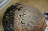
- Radiation Dermatitis
- Description:
- Radiation dermatitis - scaly pigmented scalp
Radiation dermatitis is an acute or chronic inflammation of the skin caused by exposure to ionizing radiation, as in cancer radiation therapy. Symptoms, which may not appear until 3 weeks after exposure, include redness, blistering, and sloughing of the skin. In severe cases the condition can progress to scarring, fibrosis, and atrophy. There may also be changes in skin pigmentation. Also called radiodermatitis.
- Keyword:
- Skin, radiation therapy, radiation exposure, Radiodermatitis
- Subject:
- Dermatitis, Radiation Injuries, Wounds and Injuries, Skin and Connective Tissue Diseases, Skin Diseases
- Creator:
- Dr. P.N. Girish, MBBS, MD, DDV, DNB
AJ Institute of Medical Science
- Publisher:
- AJ Institute of Medical Science
- Language:
- English
- Copyright Holder:
- AJ Institute of Medical Science
- Rights:
- http://www.i-human.com/service-agreement-print
- Resource Type:
- Photo
- Identifier:
- 2007
- Title:
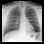
- X-ray (chest), AP, Adult Male, Thoracic Bullet, Annotated
- Description:
- CXR AP and Lat - Adult male, thoracic bullet
1. Foreign body, more radio opaque than bone, consistent with a metallic object such as a bullet. It is unclear if this object is located within the thoracic or the abdominal cavity. Addition of the lateral view shows the bullet lies either deep within the posterior chest cavity or in the paraspinous musculature.
2. Curved, meniscus-shaped blunting of the left costophrenic angle consistent with pleural effusion. If this study was obtained in the setting of acute trauma then this finding would most likely represent an acute hemothorax and indicate the placement of a chest tube during ED stabilization. Also a bedside ultrasound to check for pericardial effusion would be very useful and potentially lifesaving. Note there is no evidence of pneumothorax: lung markings can be seen all the way to the periphery of both lungs. Also, if there were a hemo-pneumothorax on the left the pleural fluid would layer with an air-fluid level, not be meniscus-shaped.
- Keyword:
- Diagnostic X-Ray, Radiography, Thoracic, Bullet, Roentgenography, Wounds and Injuries, Radiology, Diagnostic X-Ray, Diagnostic X-Ray Radiology, Foreign Bodies, X-Ray, Diagnostic, Diagnosis, X-Ray Radiology, Diagnostic
- Subject:
- Diagnostic Techniques and Procedures, Multimodal Imaging, Foreign Bodies, Diagnostic Imaging, Wounds and Injuries
- Creator:
- Anurag Agarwal, MD, Radiologist, NBE (radiograph)Lars Ensign, MD (annotations)
- Publisher:
- NBE
- Language:
- English
- Copyright Holder:
- Anurag Agarwal, MD
- Rights:
- http://www.i-human.com/service-agreement-print
- Resource Type:
- Medical Imaging
- Identifier:
- 1383
