Search
« Previous |
1,871 - 1,880 of 4,888
|
Next »
Search Results

- Title:
- CT (chest), Pulmonary Edema
- Description:
- CT shows symmetric pulmonary edema and enlarged right atrium
- Keyword:
- Pulmonary Edemas, CAT Scan, X Ray, X-Ray Computer Assisted Tomography, Computerized Tomography, X-Ray, Wet Lung, Computed Tomography, X-Ray, CT X Ray, Diagnosis, Edema, Pulmonary, Computed X Ray Tomography, Electron Beam Tomography, Tomodensitometry, Electron Beam Computed Tomography, CAT Scan, X-Ray, Tomography, X-Ray Computer Assisted, Tomography, Transmission Computed, Cine-CT, X Ray Tomography, Computed, X Ray Computerized Tomography, Tomography, Xray Computed, X-Ray Tomography, Computed, Edemas, Pulmonary, X-Ray Computerized Axial Tomography, Tomography, X-Ray Computerized, Tomography, X-Ray Computerized Axial, Computerized Tomography, X Ray, Tomography, X Ray Computed, CT Scan, X-Ray
- Subject:
- Lung Diseases, Multimodal Imaging, Diagnostic Imaging, Diagnostic Techniques and Procedures, Tomography, X-Ray Computed, Respiratory Tract Diseases, Pulmonary Edema
- Creator:
- Paul Kent, M.D.Rush Medical College
- Publisher:
- Rush Medical College
- Language:
- English
- Copyright Holder:
- Rush Medical College
- Rights:
- http://www.i-human.com/service-agreement-print
- Resource Type:
- Medical Imaging
- Identifier:
- 1797
- Title:
- Transthoracic Echocardiogram (TTE), Atrial Septal Defect (ASD)
- Description:
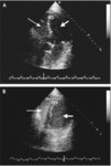
- The image in Panel A shows minimal right-to-left shunting through the patent foramen ovale (arrow). It was obtained with the patient in the supine position.
The image in Panel B shows increased right-to-left shunting (arrow). It was obtained with the patient in the sitting position. LA denotes left atrium, RA right atrium, and Ao aorta.
- Keyword:
- Echocardiography, M-Mode, Transthoracic Echocardiography, Echocardiography, 2-D, Cross-Sectional Echocardiography, 2D Echocardiography, Atrial Septal Defects, Echocardiography, Cross-Sectional, Heart, Echocardiography, Two-Dimensional, Persistent Ostium Primum, Diagnosis, Echocardiography, 2D, Echocardiography, Contrast, Atrial Septal Defect, Echocardiography, Transthoracic, Ostium Secundum Atrial Septal Defect, 2-D Echocardiography, M-Mode Echocardiography, Two-Dimensional Echocardiography, Contrast Echocardiography
- Subject:
- Cardiac Imaging Techniques, Multimodal Imaging, Echocardiography, Heart Defects, Congenital, Cardiovascular Diseases, Diagnostic Imaging, Cardiovascular Abnormalities, Diagnostic Techniques and Procedures, Heart Septal Defects
- Creator:
- Paul Kent, M.D. Rush Medical College
- Publisher:
- i-Human Patients, Inc.
- Language:
- English
- Copyright Holder:
- Paul Kent, M.D.
- Rights:
- http://www.i-human.com/service-agreement-print
- Resource Type:
- Medical Imaging
- Identifier:
- 1802
- Title:
- Transthoracic Echocardiogram (TTE), Atrial Septal Defect (ASD)
- Description:
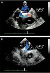
- Note the passage of bubbles from the RA to the LA and flow (blue towards the transducer, red away) on the Doppler echo.
- Keyword:
- 2D Echocardiography, Atrial Septal Defect, Persistent Ostium Primum, Echocardiography, 2-D, Echocardiography, Cross-Sectional, Ostium Secundum Atrial Septal Defect, M-Mode Echocardiography, Two-Dimensional Echocardiography, 2-D Echocardiography, Echocardiography, M-Mode, Echocardiography, Two-Dimensional, Echocardiography, 2D, Transthoracic Echocardiography, Cross-Sectional Echocardiography, Echocardiography, Transthoracic, Heart, Diagnosis, Atrial Septal Defects, Contrast Echocardiography, Echocardiography, Contrast
- Subject:
- Echocardiography, Diagnostic Techniques and Procedures, Diagnostic Imaging, Cardiac Imaging Techniques, Multimodal Imaging, Heart Septal Defects, Cardiovascular Abnormalities, Heart Defects, Congenital, Cardiovascular Diseases
- Creator:
- Paul Kent, M.D.
- Publisher:
- i-Human Patients, Inc.
- Language:
- English
- Copyright Holder:
- Paul Kent, M.D.
- Rights:
- http://www.i-human.com/service-agreement-print
- Resource Type:
- Medical Imaging
- Identifier:
- 1798
- Title:
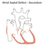
- Atrial septal defect types - ostium secundum defect
- Description:
- ASD primum
ASD secundum
ASD sinus venosus
- Keyword:
- atrial septal defect, congenital heart defect, Heart Defects, Congenital, Ostium secundum defect, right to left shunt, Heart Septal Defects, Atrial, Lutembacher Syndrome
- Subject:
- Endocardial Cushions
- Creator:
- i-Human Patients, Inc.
- Publisher:
- i-Human Patients, Inc.
- Language:
- English
- Copyright Holder:
- i-Human Patients, Inc.
- Rights:
- All rights reserved
- Resource Type:
- Illustration
- Identifier:
- 1803
- Title:
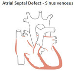
- Atrial septal defect types - sinus venosus defect
- Description:
- ASD primum
ASD secundum
ASD sinus venosus
- Keyword:
- Right To Left Shunt, Heart Defects, Congenital, Atrial Septal Defect, Heart Septal Defects, Atrial, Atrial Septal Defect Sinus Venosus, Sinus Venosus
- Subject:
- Endocardial Cushions
- Creator:
- i-Human Patients, Inc.
- Publisher:
- i-Human Patients, Inc.
- Language:
- English
- Copyright Holder:
- i-Human Patients, Inc.
- Rights:
- All rights reserved
- Resource Type:
- Illustration
- Identifier:
- 1803
- Title:
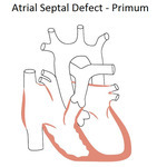
- Atrial septal defect types - ostium primum defect
- Description:
- ASD primum
ASD secundum
ASD sinus venosus
- Keyword:
- atrial septal defect, Right to Left Shunt, Heart Defects, Congenital, Congenital Heart Defect, Heart Septal Defects, Atrial, Ostium primum defect
- Subject:
- Endocardial Cushions
- Creator:
- i-Human Patients, Inc.
- Publisher:
- i-Human Patients, Inc.
- Language:
- English
- Copyright Holder:
- i-Human Patients, Inc.
- Rights:
- All rights reserved
- Resource Type:
- Illustration
- Identifier:
- 1803
- Title:
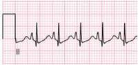
- 3 Lead ECG: Right Ventricular Hypertrophy (RVH)
- Description:
- Lead V1 show rSR' (with no widening of QRS complex), this is one of several ways RVH is exhibited in ECG, other forms of RVH manifestation on ECG include:
Tall R in V1 + deep S in V6
Pure R wave in V1
qR wave in V1
Abnormal direction of T wave in right chest leads (normal T waves are downward in right chest leads in children; therefore upright T waves in V1-3 could indicate RVH.
Lead II show tall P wave indicating right atrial enlargement (taller than 2 mm).
- Keyword:
- Pulmonary Embolism, Heart Ventricles, ECG, Right Ventricular Hypertrophy, EKG, Hypertrophies, Right Ventricular, Pulmonary Circulation, Right Ventricular Hypertrophies, Right Atrial Enlargement, Heart, Pulmonary Hypertension, Ventricular Hypertrophies, Right, Diagnosis, Electrocardiogram, Electrocardiograph, Ventricular Hypertrophy, Right
- Subject:
- Hypertrophy, Right Ventricular, Hypertension, Pulmonary, Hypertrophy, Embolism, Lung Diseases, Cardiomegaly, Diagnostic Techniques and Procedures, Diagnostic Techniques, Cardiovascular, Respiratory Tract Diseases, Electrocardiography, Vascular Resistance
- Creator:
- Paul Kent, MDRush Medical College
- Publisher:
- Rush Medical College
- Language:
- English
- Copyright Holder:
- Rush Medical College
- Rights:
- http://www.i-human.com/service-agreement-print
- Resource Type:
- Chart/Diagram
- Identifier:
- 1799
- Title:
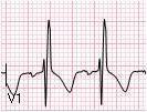
- 3 Lead ECG - Right Ventricular Hypertrophy (RVH)
- Description:
- Lead V1 show rSR' (with no widening of QRS complex), this is one of several ways RVH is exhibited in ECG, other forms of RVH manifestation on ECG include:
Tall R in V1 + deep S in V6
Pure R wave in V1
qR wave in V1
Abnormal direction of T wave in right chest leads (normal T waves are downward in right chest leads in children; therefore upright T waves in V1-3 could indicate RVH.
Lead II show tall P wave indicating right atrial enlargement (taller than 2 mm).
- Keyword:
- Electrocardiograph, Pulmonary Hypertension, EKG, ECG, Right Atrial Enlargement, Ventricular Hypertrophies, Right, Heart Ventricles, Right Ventricular Hypertrophies, Electrocardiogram, Right Ventricular Hypertrophy, Ventricular Hypertrophy, Right, Diagnosis, Hypertrophies, Right Ventricular, Heart, Pulmonary Circulation, Pulmonary Embolism
- Subject:
- Diagnostic Techniques and Procedures, Diagnostic Techniques, Cardiovascular, Embolism, Cardiomegaly, Electrocardiography, Hypertrophy, Right Ventricular, Hypertension, Pulmonary, Hypertrophy, Vascular Resistance, Respiratory Tract Diseases, Lung Diseases
- Creator:
- Paul Kent, MD
Rush Medical College
- Publisher:
- Rush Medical College
- Language:
- English
- Copyright Holder:
- Rush Medical College
- Rights:
- http://www.i-human.com/service-agreement-print
- Resource Type:
- Chart/Diagram
- Identifier:
- 1799
- Title:
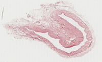
- External Jugular Vein
- Description:
- System: Cardiovascular
Organ: External jugular vein
Disease process: Normal
Species: Human
Highest magnification: 20x
Stain: H&E
Note that there is no external elastic membrane, that the middle coat is thin and the smooth muscle is loosely organized.
- Keyword:
- vein, morphology, blod vessels, Anatomy & histology, external jugular vein, vascular system, anatomy, histology
- Subject:
- Veins, Blood Vessels, Cardiovascular System, Histological Techniques, Staining and Labeling, Histocytological Preparation Techniques, Jugular Veins
- Creator:
- John R. Cotter, PhD Department of Pathology and Anatomical Sciences University at Buffalo School of Medicine
- Publisher:
- University at Buffalo School of Medicine
- Language:
- English
- Rights:
- All rights reserved
- Resource Type:
- Slide
- Identifier:
- 1790

- Title:
- Vena Cava, Monkey
- Description:
- H&E & Kornhauser quadruple
- Keyword:
- cavae, venae, vena cava, monkey, morphology, histology, monkeys, anatomy, Anatomy & histology
- Subject:
- Primates, Vena Cava, Inferior, Staining and Labeling, Histocytological Preparation Techniques, Histological Techniques, Cardiovascular System, Venae Cavae, Blood Vessels, Vena Cava, Superior, Veins
- Creator:
- John Cotter, PhD
Department of Pathology and Anatomical Sciences
University at Buffalo School of Medicine
- Publisher:
- University at Buffalo School of Medicine
- Language:
- English
- Copyright Holder:
- University at Buffalo School of Medicine
- Rights:
- http://www.i-human.com/service-agreement-print
- Resource Type:
- Slide
- Identifier:
- 1791
