Search
« Previous |
1 - 10 of 751
|
Next »
Search Results
Select an image to start the slideshow
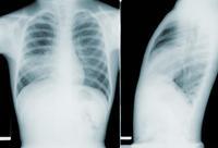
X-ray (chest), Pneumonia RLL
1 of 10

UltrasoundOvarianCyst
2 of 10
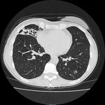
Chest CT, Mid-lung nodular bronchiectasis
3 of 10
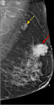
MRI Left Breast
4 of 10
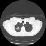
CT (Lung), Right Upper Lobe Cavity with Surrounding Infiltrate
5 of 10
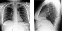
X-ray (chest), PA and Lateral, Adult Female, Normal
6 of 10
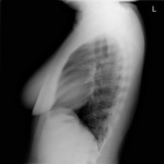
X-ray (chest), Lateral, Female, Hyperinflation
7 of 10
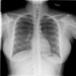
X-ray (chest), PA, Female, Hyperinflation
8 of 10
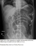
CT (abdomen), (lateral supine), Small Bowel Obstruction (annotated)
9 of 10
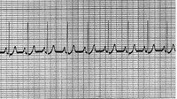
3 Lead ECG: Normal Sinus Rhythm (NSR) ~90 bpm
10 of 10
