Search
« Previous |
1 - 10 of 15
|
Next »
Search Results
Select an image to start the slideshow
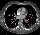
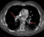
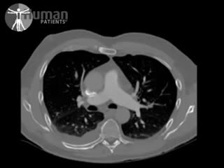
CT pulmonary angiography (CTPA) 5, Pulmonary embolism (PE)
1 of 10
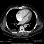
CT pulmonary angiography (CTPA) 6, Pulmonary embolism (PE)
2 of 10
CT pulmonary angiography (CTPA) 2, Pulmonary embolism (PE)
3 of 10
CT pulmonary angiography (CTPA) 3, Pulmonary embolism (PE)
4 of 10
CT pulmonary angiography (CTPA) 4, Pulmonary embolism (PE)
5 of 10
CT pulmonary angiography (CTPA) 1, Pulmonary embolism (PE)
6 of 10
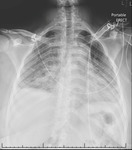
X-ray (chest), Acute Pulmonary Embolism
7 of 10
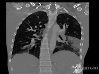
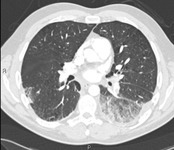
CT (chest), Pulmonary Embolism with Infarction, Anterior to Posterior
8 of 10
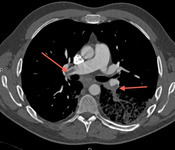
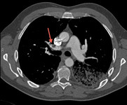
CT (chest), (axial), Pulmonary Embolism
9 of 10
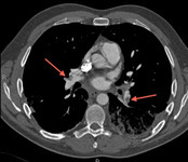
CT (chest), (axial), Adult Male Pulmonary Embolism with Right Heart Strain
10 of 10
