Search
« Previous |
101 - 110 of 1,001
|
Next »
Search Results
Select an image to start the slideshow

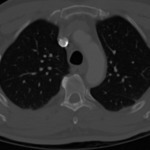
CT (chest), Normal
1 of 10
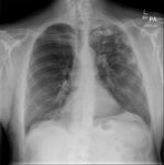
X-ray (chest), PA, Adult Male, Tuberculosis
2 of 10
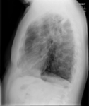
X-ray (chest), Lateral, Adult Male, Tuberculosis
3 of 10
X-ray (chest), Adult Male, Tuberculosis
4 of 10
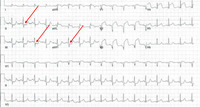
12 Lead ECG Inferior lead ST depression
5 of 10
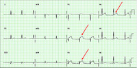
12 Lead ECG: Anterior STEMI
6 of 10
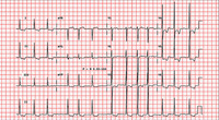
12 Lead ECG Atrial Fibrillation with left ventricular hypertrophy (LVH)
7 of 10

12 Lead ECG: Atrial Fibrillation
8 of 10
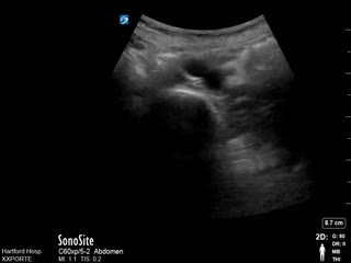
Ultrasound (abdomen), Abdominal Aortic Aneurysm (AAA)
9 of 10
layer09_24861.dcm
10 of 10
