Search
« Previous |
31 - 40 of 1,547
|
Next »
Search Results
Select an image to start the slideshow
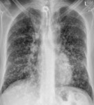
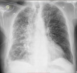
Miliary Tuberculosis (TB), Adult Male, CXR PA
2 of 10
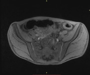
MRI: Ulcerative Colitis
3 of 10
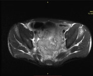
MRI: Ulcerative Colitis
4 of 10
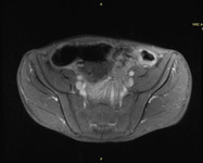
MRI: Ulcerative Colitis
5 of 10
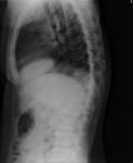
Xray Spine, Pott's Disease
6 of 10
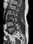
MRI Lumbar Spine - Metastases
7 of 10
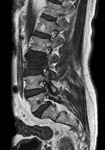
MRI Lumbar Spine - Metastases
8 of 10
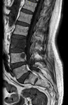
MRI Lumbar Spine - Metastases
9 of 10
CXR AP-L Interstitial Lung Disease
10 of 10
