×
You need to sign in or sign up before continuing.
Search
« Previous |
491 - 500 of 848
|
Next »
Search Results
-
| show more |
| Title: |
Epididymis |
| Depositor: |
batchuser@i-human.com |
| Creator: |
Rush Medical College |
| Histology: Epididymis |
| Keywords: |
Anatomy & histology, Epidydymis, morphology, male reproductive system, anatomy, histology, Genitalia, Male |
| Date Uploaded: |
07/29/2012 |
-
| show more |
| Title: |
Eye |
| Depositor: |
batchuser@i-human.com |
| Creator: |
Rush Medical College |
| System: Neurologic
Organ: Eye
Diagnosis: Normal
Species: Human
Magnification: 20x
Stain: |
| Keywords: |
anatomy, morphology, Anatomy & histology, histology |
| Date Uploaded: |
07/29/2012 |
-
| show more |
| Title: |
Eye, Retina and Optic Disc |
| Depositor: |
batchuser@i-human.com |
| Creator: |
Rush Medical College |
| System: Nervous
Organ: Eye retina and optic disc
Diagnosis: Normal
Species: Human
Magnification: 20x
Stain: |
| Keywords: |
morphology, retina, Anatomy & histology, eye, anatomy, histology, optic disc |
| Date Uploaded: |
07/29/2012 |
-
| show more |
| Title: |
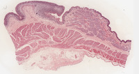
Gastro-esophageal Junction |
| Depositor: |
batchuser@i-human.com |
| Creator: |
Rush Medical College |
| System: Gastrointestinal
Organ: Esophageal cardiac junction
Disease process: Normal
Species: Human
Highest magnification: 20x
Stain: H&E
Note the epithelial transition from stratified squamous non-keratinized (esophagus) to simple columnar (gastric). There are two sets of glands: esophageal glands (in the submucosa) vs. cardiac gastric glands (in the mucosa).
In the muscularis layer the “sphincter” is an expansion of the middle oblique layer in the gastric muscularis which forms an incomplete sphincter, i.e., “sling” or valve + skeletal muscle (crura of diaphragm).
The cells that line the gastric glands in the cardiac region of the stomach: they are all mucous producing cells. The luminal surface and the gastric pits are also lined by surface mucus cells. |
| Keywords: |
histology, small intestine, Anatomy & histology, digestive system, gastro-esophageal junction, morphology, anatomy, esophageal-cardiac junction |
| Date Uploaded: |
07/29/2012 |
-
| show more |
| Title: |
Kidney |
| Depositor: |
batchuser@i-human.com |
| Creator: |
Rush Medical College |
| System: Urologic
Organ: Kidney
Disease process: Normal
Species: Rat
Stain: H&E
Highest magnification: 20x
Coronal section with flattened space of the renal calyx and the loose connective tissue and vessels at the hilus. |
| Keywords: |
anatomy, histology, kidney, kidneys, morphology, Anatomy & histology |
| Date Uploaded: |
07/29/2012 |
-
| show more |
| Title: |
Eye, Retina |
| Depositor: |
batchuser@i-human.com |
| Creator: |
Rush Medical College |
| System: Nervous
Organ: Retina
Diagnosis: Normal
Magnification: 20x
Stain: H&E |
| Keywords: |
morphology, Anatomy & histology, anatomy, histology, eye |
| Date Uploaded: |
07/29/2012 |
-
| show more |
| Title: |
Eyelid |
| Depositor: |
batchuser@i-human.com |
| Creator: |
Rush Medical College |
| System: Integumentary
Organ: Eye lid
Diagnosis: Normal
Stain:
Magnification: |
| Keywords: |
histology, anatomy, eyelid, morphology, eye, Anatomy & histology |
| Date Uploaded: |
07/29/2012 |
-
| show more |
| Title: |
Eye, Anterior Segment |
| Depositor: |
batchuser@i-human.com |
| Creator: |
Rush Medical College |
| Histology: Eye, anterior segment |
| Keywords: |
histology, anatomy, Anatomy & histology, anterior segment of eye, morphology |
| Date Uploaded: |
07/29/2012 |
-
| show more |
| Title: |
Gall Bladder |
| Depositor: |
batchuser@i-human.com |
| Creator: |
Rush Medical College |
| System: Gastrointestinal
Organ: Gallbladder
Disease process: Normal
Species: Human
Stain: H&E
Highest magnification: 20x |
| Keywords: |
gall bladder, bile, Anatomy & histology, anatomy, histology, morphology |
| Date Uploaded: |
07/29/2012 |
-
| show more |
| Title: |
Bone (ground) |
| Depositor: |
batchuser@i-human.com |
| Creator: |
Rush Medical College |
| System: Skeletal
Organ: Bone, ground
Diagnosis: Normal
Disease process: Normal
Species: Human
Highest magnification: 20x
Stain: ? |
| Keywords: |
anatomy, bone, histology, Musculoskeletal Diseases, morphology, Anatomy & histology, ground bone |
| Date Uploaded: |
07/29/2012 |
