Search
« Previous |
1 - 10 of 2,494
|
Next »
Search Results

- Title:
- UltrasoundOvarianCyst
- Description:
- thin-walled hypoechoic ovarian cyst
- Subject:
- Ultrasonography, Diagnostic Imaging, Diagnostic Techniques and Procedures, Diagnosis, pelvis
- Creator:
- Courtney Anderson
- Rights:
- http://creativecommons.org/licenses/by/3.0/us/
- Resource Type:
- Medical Imaging
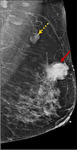
- Title:
- MRI Left Breast
- Description:
- MRI of the left breast mass revealed 4-cm oval mass (red arrow) and an enlarged axillary lymph node (yellow arrow)
With permission from Sharma S, Vicenty-Latorre FG, Elsherif S, Sharma S.
- Keyword:
- Neoplasm, Cancer, Breast cancer
- Subject:
- Multimodal Imaging, Diagnostic Imaging, Diagnostic Techniques and Procedures, Diagnosis
- Creator:
- Sharma S, Vicenty-Latorre FG, Elsherif S, Sharma S
- Publisher:
- Sharma S, Vicenty-Latorre FG, Elsherif S, Sharma S. Role of MRI in breast cancer staging: a case-based review. Cureus. 2021;13(12):e20752.8
- Copyright Holder:
- Sharma S, Vicenty-Latorre FG, Elsherif S, Sharma S
- Rights:
- https://creativecommons.org/licenses/by/3.0
- Resource Type:
- Medical Imaging
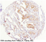
- Title:
- Invasive Breast Cancer, IHC 2+ (equivocal)
- Description:
- The revised definition of IHC 2+ (equivocal) is invasive breast cancer with “weak to moderate complete membrane staining observed in >10% of tumor cells.”
- Keyword:
- Cancer of Breast, Breast Cancer, Neoplasms, Breast, Malignant Neoplasm of Breast, Breast Tumor, Breast Carcinoma
- Subject:
- Multimodal Imaging, Breast Neoplasms, Diagnostic Imaging, Diagnostic Techniques and Procedures, Diagnosis, Neoplasms by Site, Neoplasms
- Creator:
- Image courtesy of Tiffany A. Traina, MD
- Publisher:
- i-Human Patients, Inc.
- Copyright Holder:
- Tiffany A. Traina, MD
- Rights:
- http://www.i-human.com/service-agreement-print
- Resource Type:
- Slide
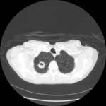
- Title:
- CT (Lung), Right Upper Lobe Cavity with Surrounding Infiltrate
- Description:
- CT scan of lung showing right upper lobe cavity with surrounding infiltrate
- Keyword:
- CAT Scan, X-ray, CT Scan, Lung CT, CT Lung
- Subject:
- Diagnostic Techniques and Procedures, Multimodal Imaging, Lung Diseases, Diagnosis, Diagnostic Imaging, Respiratory Tract Diseases
- Creator:
- Kevin Winthrop, MD, MPH
- Publisher:
- i-Human Patients, Inc.
- Copyright Holder:
- Kevin Winthrop, MD, MPH
- Rights:
- http://www.i-human.com/service-agreement-print
- Resource Type:
- Medical Imaging
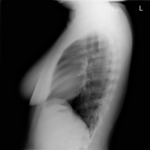
- Title:
- X-ray (chest), Lateral, Female, Hyperinflation
- Description:
- Mildly hyperinflated lungs with flattened posterior diaphragm. No alveolar consolidation, no findings of pleural effusion or pulmonary edema. Heart size within normal limits. No pneumothorax. Mildly hyperinflated lungs, air trapping versus inspiratory.
- Keyword:
- Chest x-ray, Diagnosis, Chest xray, Hyperinflation, Lung hyperinflation, Hyperinflated lungs
- Subject:
- Diagnostic Imaging, Diagnostic Techniques and Procedures, Diagnosis
- Creator:
- Kohli MD, Rosenman M, Indiana University
- Publisher:
- i-Human Patients, Inc.
- Copyright Holder:
- Kohli MD, Rosenman M
- Rights:
- http://creativecommons.org/publicdomain/mark/1.0/
- Resource Type:
- Medical Imaging
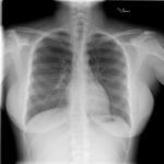
- Title:
- X-ray (chest), PA, Female, Hyperinflation
- Description:
- Mildly hyperinflated lungs with flattened posterior diaphragm. No alveolar consolidation, no findings of pleural effusion or pulmonary edema. Heart size within normal limits. No pneumothorax. Mildly hyperinflated lungs, air trapping versus inspiratory.
- Keyword:
- Chest x-ray, Diagnosis, Chest xray, Hyperinflation, Lung hyperinflation, Hyperinflated lungs, Roentgenography, X-Ray Radiology, Diagnostic, Diagnostic X-Ray Radiology
- Subject:
- Diagnostic Imaging, Diagnostic Techniques and Procedures, Diagnosis
- Creator:
- Kohli MD, Rosenman M, Indiana University
- Publisher:
- i-Human Patients, Inc.
- Copyright Holder:
- Kohli MD, Rosenman M, Indiana University
- Rights:
- http://creativecommons.org/publicdomain/mark/1.0/
- Resource Type:
- Medical Imaging
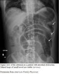
- Title:
- CT (abdomen), (lateral supine), Small Bowel Obstruction (annotated)
- Description:
- Supine view of the abdomen in a patient with intestinal obstruction. Dilated loops of small bowel are visible (arrows).
- Keyword:
- Intestinal Obstruction, X Ray Computerized Tomography, Computed Tomography, X-Ray, Tomodensitometry, Electron Beam Tomography, Tomography, X-Ray Computer Assisted, Tomography, X-Ray Computerized, X-Ray Computerized Axial Tomography, CT Scan, X-Ray, Electron Beam Computed Tomography, X-Ray Tomography, Computed, Tomography, X-Ray Computerized Axial, Computerized Tomography, X Ray, CAT Scan, X-Ray, X Ray Tomography, Computed, Gastrointestinal Diseases, X-Ray Computer Assisted Tomography, Tomography, X Ray Computed, Digestive System Diseases, Tomography, Transmission Computed, Small Bowel Obstruction, Bowel, Diagnosis, CT X Ray, Computerized Tomography, X-Ray, Intestinal Diseases, Cine-CT, Computed X Ray Tomography, Obstruction, SBO, Tomography, Xray Computed, CAT Scan, X Ray
- Subject:
- Gastrointestinal Diseases, Intestinal Diseases, Multimodal Imaging, Intestinal Obstruction, Tomography, X-Ray Computed, Diagnostic Imaging, Diagnostic Techniques and Procedures, Digestive System Diseases, Diagnosis, Image Interpretation, Computer-Assisted
- Creator:
- diana@i-human.com
- Publisher:
- i-Human Patients, Inc.
- Copyright Holder:
- American Family Physician
- Rights:
- http://creativecommons.org/licenses/by/3.0/us/
- Resource Type:
- Medical Imaging
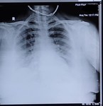
- Title:
- X-ray (chest), PA, COVID-19
- Description:
- X-ray (chest), PA, COVID-19. X-ray (chest), PA, COVID-19. Oxygen therapy, no evidence of pneumonia.
- Keyword:
- Lung Inflammation, X-Ray, Diagnostic, Respiratory Disease, Lung Disease, Respiratory Infection, Viral Disease, Corona Virus
- Subject:
- Radiography, Thoracic, Lung Diseases, Respiratory Tract Diseases, Diagnostic Imaging, Radiography
- Creator:
- Neethi Chandra
- Rights:
- http://www.i-human.com/service-agreement-print, https://creativecommons.org/licenses/by-sa/3.0
- Resource Type:
- Medical Imaging
- Title:
- X-ray (chest), PA, COVID-19
- Description:
- X-ray (chest), PA, COVID-19. X-ray (chest), PA, COVID-19. Oxygen therapy, no evidence of pneumonia.
- Keyword:
- Lung Inflammation, X-Ray, Diagnostic, Respiratory Disease, Lung Disease, Respiratory Infection, Viral Disease, Corona Virus
- Subject:
- Radiography, Thoracic, Lung Diseases, Respiratory Tract Diseases, Radiography, Diagnostic Imaging
- Creator:
- Neethi Chandra
- Rights:
- https://creativecommons.org/licenses/by-sa/3.0
- Resource Type:
- Medical Imaging
- Title:
- X-ray (chest), PA, COVID-19
- Description:
- X-ray (chest), PA, COVID-19. X-ray (chest), PA, COVID-19. Oxygen therapy, no evidence of pneumonia.
- Keyword:
- Lung Inflammation, X-Ray, Diagnostic, Respiratory Disease, Lung Disease, Respiratory Infection, Viral Disease, Corona Virus
- Subject:
- Radiography, Thoracic, Lung Diseases, Respiratory Tract Diseases, Diagnostic Imaging, Radiography
- Creator:
- Neethi Chandra
- Rights:
- https://creativecommons.org/licenses/by-sa/3.0
- Resource Type:
- Medical Imaging
