Search
« Previous |
31 - 40 of 313
|
Next »
Search Results
Select an image to start the slideshow
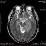
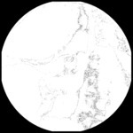
Temporal Neoplasm
1 of 10
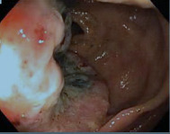
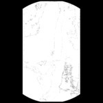
Colon Cancer, Endoscopy
2 of 10

PET-CT (whole body), Esophageal Cancer, Image 2
3 of 10
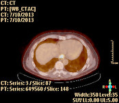
PET-CT (whole body), Esophageal Cancer, Image 1
4 of 10
Esophageal Cancer, Fluoro Esophagogram, Image 1
5 of 10
Esophageal Cancer, Fluoro Esophagogram, Image 3
6 of 10
Esophageal Cancer, Fluoro Esophagogram, Image 2
7 of 10
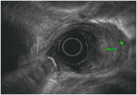
Ultrasound (esophagus), Esophageal Mass
8 of 10
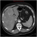
CT Abdomen (axial), Liver Metastases
9 of 10
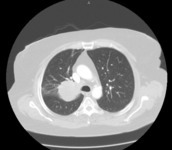
CT (chest), Lung Mass, Upper Right Lobe
10 of 10
