×
You need to sign in or sign up before continuing.
Search
Search Results
-
| show more |
| Title: |
Heart cross section great vessels |
| Depositor: |
batchuser@i-human.com |
| Creator: |
i-Human Patients, Inc. |
| Heart illustration in cross section with great vessels, ABCs |
| Keywords: |
Vena Cava, Superior, Heart, arch of the aorta, Heart Ventricles, Vena Cava, Inferior, heart chambers, inferior vena cava, superior vena cava, pulmonary artery, illustration |
| Is part of: |
Heart cross section great vessels |
| Date Uploaded: |
04/22/2016 |
-
| show more |
| Title: |
Great Vessels And Heart |
| Depositor: |
batchuser@i-human.com |
| Creator: |
Kristina DeRycke
i-Human Patients, Inc. |
| Illustration of heart and the great vessels, labeled. |
| Keywords: |
Subclavian Vein, Subclavian Artery, Vena Cava, Superior, Common Carotid Artery, Pulmonary Veins, Carotid Artery, Common, Pulmonary Artery, Superior Vena Cava, Heart, Inferior Vena Cava, Vena Cava, Inferior, Cardiovascular System, Jugular Veins, Brachiocephalic Trunk |
| Date Uploaded: |
06/01/2015 |
-
| show more |
| Title: |
Respiratory System |
| Depositor: |
batchuser@i-human.com |
| Creator: |
Kristina DeRycke
i-Human Patients, Inc. |
| Anatomy of the respiratory system. |
| Keywords: |
Gas Exchange, Reperfusion, Alveoli, Pulmonary Ventilation, Ventilation/ Perfusion anatomy, Respiratory System, Respiratory Tree |
| Date Uploaded: |
05/27/2015 |
-
| show more |
| Title: |
Airway Diagram |
| Depositor: |
batchuser@i-human.com |
| Creator: |
Kristina DeRycke
i-Human Patients, Inc. |
| Airways, tracheal/bronchial, lung parenchyma |
| Keywords: |
Alveoli, Ventilation/ Perfusion anatomy, Respiratory System, Reperfusion, Pulmonary Ventilation, Gas Exchange, Respiratory Tree |
| Date Uploaded: |
05/27/2015 |
-
| show more |
| Title: |
Cardiac Great Vessels and Coronary Vessels; Labels |
| Depositor: |
batchuser@i-human.com |
| Creator: |
Laura Garrison
i-Human Patients; Inc. |
| Animation - Cardiac great vessels & coronary vessels |
| Keywords: |
Cardiovascular System, Aorta, Thoracic, Superior Vena Cava, Vena Cava, Inferior, Vena Cava, Superior, Thoracic Aorta, Coronary Sinus, Heart, Inferior Vena Cava, Pulmonary Artery, Aorta |
| Date Uploaded: |
10/07/2013 |
-
| show more |
| Title: |
Cardiac Great Vessels and Coronary Vessels; Matching Exercise |
| Depositor: |
batchuser@i-human.com |
| Creator: |
Laura Garrison
i-Human Patients; Inc. |
| Animation - Cardiac great vessels & coronary vessels |
| Keywords: |
Cardiovascular System, Vena Cava, Superior, Vena Cava, Inferior, Pulmonary Artery, Inferior Vena Cava, Aorta, Coronary Sinus, Superior Vena Cava, Heart, Aorta, Thoracic, Thoracic Aorta |
| Date Uploaded: |
10/07/2013 |
-
| show more |
| Title: |
Diagnostic algorithm: Pulmonary embolism |
| Depositor: |
batchuser@i-human.com |
| Creator: |
L.M. Tierney; M.C. Henderson |
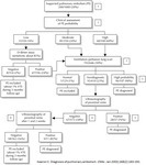
| A diagnostic algorithm for pulmonary embolism (estimated frequencies of test results and associated prevalences of pulmonary embolism for a hypothetical cohort of 1000 outpatients) [1]. If a very sensitive D-dimer assay is used, it can be the first test performed: a negative result excludes pulmonary embolism regardless of clinical assessment category and a positive test can be followed by a ventilation–perfusion scan [2]. A ventilation–perfusion scan can be performed as the initial test without using clinical assessment of the probability of pulmonary embolism as part of the diagnostic process [3]. Pulmonary angiography or helical CT may be considered if the clinical assessment of pulmonary embolism probability is low, particularly if a D-dimer test has not been done [4]. Additional testing (e.g., helical CT, bilateral venography) may be considered if overall assessment suggests a high probability of pulmonary embolism (e.g., 50%–80%), symptoms are severe or cardiopulmonary reserve is poor [5]. Venography should be considered if there is an increased risk of a false-positive ultrasound result (e.g., previous venous thromboembolism, equivocal ultrasound findings, preceding findings suggest low probability of pulmonary embolism [e.g., ≤ 10%]) [6]. It is reasonable not to repeat ultrasound testing, or to do only 1 more ultrasound after 1 week, if preceding findings suggest a low probability of pulmonary embolism (e.g., ≤ 10%) [7]. If helical CT is used in place of ventilation–perfusion lung scanning: (i) intraluminal filling defects in segmental or larger pulmonary arteries are generally diagnostic for pulmonary embolism; (ii) all other findings (i.e., a normal CT scan or intraluminal filling defects confined to the subsegmental pulmonary arteries) are nondiagnostic and can be managed as shown for a nondiagnostic lung scan. |
| Keywords: |
Embolism, Pulmonary Thromboembolism, Thromboembolism, Pulmonary, Embolism, Pulmonary
Pulmonary Thromboembolisms, Embolisms, Pulmonary, Thromboembolisms, Pulmonary, Vascular Diseases, Pulmonary Embolisms |
| Date Uploaded: |
06/23/2013 |
-
| show more |
| Title: |
Heart cross section, matching |
| Depositor: |
batchuser@i-human.com |
| Creator: |
i-Human Patients, Inc. |
| Heart illustration in cross section with great vessels, ABCs |
| Keywords: |
superior vena cava, arch of the aorta, inferior vena cava, Heart, heart chambers, Heart Ventricles, Vena Cava, Superior, Vena Cava, Inferior, pulmonary artery, illustration |
| Date Uploaded: |
01/19/2013 |
-
| show more |
| Title: |
Flow of blood through the heart |
| Depositor: |
batchuser@i-human.com |
| Creator: |
|
| Heart diagram showing normal flow of blood through atria and ventricles |
| Keywords: |
inferior vena cava, Heart, Vena Cava, Inferior, heart chambers, Heart Ventricles, superior vena cava, Vena Cava, Superior, illustration, arch of the aorta, pulmonary artery |
| Date Uploaded: |
01/19/2013 |
