Search
« Previous |
101 - 110 of 862
|
Next »
Search Results
Select an image to start the slideshow
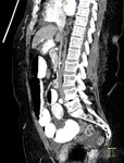
CT Abdomen (sagittal), Small Bowel Obstruction
1 of 10
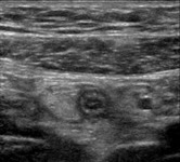
Ultrasound (abdomen), Acute Appendicitis
2 of 10
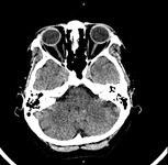
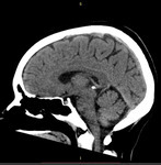
CT (brain), Normal
3 of 10
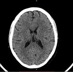
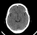
CT (brain), Normal
4 of 10
CT (brain), Normal
5 of 10
CT (brain), Normal
6 of 10
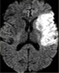
MRI (brain), Diffusion-Weighted Imaging, Left MCA Infarction
7 of 10
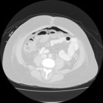
CT (abdomen), Free Intraperitoneal Air
8 of 10

Abdominal Pain 5. Right Upper Quadrant Pain Differential Diagnosis and Approach
9 of 10

Abdominal Pain 4. Epigastric Pain PUD and Pancreatitis
10 of 10
