×
You need to sign in or sign up before continuing.
Search
« Previous |
2,541 - 2,550 of 6,265
|
Next »
Search Results
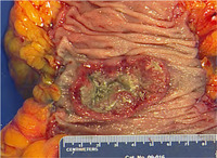
- Title:
- Colon, Adenocarcinoma
- Description:
- Colon cancer in muscularis
Gross anatomy
- Keyword:
- Colon, Anatomy, Neoplasms by Histologic Type, Neoplasms, muscularis, Carcinoma, colon cancer, Adenocarcinoma, Natural Science Disciplines
- Subject:
- Neoplasms, Glandular and Epithelial, Biological Science Disciplines
- Creator:
- Elizabeth Baker M.D., MHPEAssociate Professor of Internal MedicineAssistant Dean of Clinical EducationRush Medical College
- Publisher:
- Rush Medical College
- Language:
- English
- Copyright Holder:
- Rush Medical College
- Rights:
- http://www.i-human.com/service-agreement-print
- Resource Type:
- Photograph
- Identifier:
- 2128
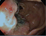
- Title:
- Colon Cancer, Endoscopy
- Description:
- Endoscopy: Colon cancer
Cancer of the colon is the disease characterized by the development of malignant cells in the lining or epithelium of the first and longest portion of the large intestine. Malignant cells have lost normal control mechanisms governing growth. These cells may invade surrounding local tissue, or they may spread throughout the body and invade other organ systems.
- Keyword:
- Diagnosis, Colon, Minimally Invasive Surgical Procedures, malignant, Digestive System Neoplasms, Colonic Neoplasms, colon cancer, Neoplasms
- Subject:
- Gastrointestinal Diseases, Colonic Neoplasms, Diagnostic Techniques, Surgical, Neoplasms by Site, Intestinal Diseases, Gastrointestinal Neoplasms, Biopsy, Diagnostic Techniques and Procedures, Surgical Procedures, Operative, Endoscopy
- Creator:
- Elizabeth Baker M.D., MHPE
Associate Professor of Internal Medicine Assistant
Dean of Clinical Education
Rush Medical College
- Publisher:
- Rush Medical College
- Language:
- English
- Copyright Holder:
- Rush Medical College
- Rights:
- http://www.i-human.com/service-agreement-print
- Resource Type:
- Photo
- Identifier:
- 2130
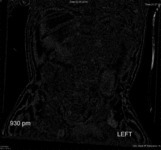
- Title:
- X-ray (abdomen), intusseption
- Description:
- X-Ray Abdomen/ KUB 1 view, 2 yo F, all
- Keyword:
- Intususception, Radiography, Diagnostic X-Ray, Diagnosis, Radiology, Diagnostic X-Ray, X-Ray, Diagnostic, Roentgenography, X-Ray Radiology, Diagnostic, Intussusception, Invagination, Intestinal, Diagnostic X-Ray Radiology
- Subject:
- Intussusception, Multimodal Imaging, Intestinal Diseases, Gastrointestinal Diseases, Diagnostic Imaging, Diagnostic Techniques and Procedures, Intestinal Obstruction, Digestive System Diseases
- Creator:
- Rush University Medical Center
- Contributor:
- i-Human-Rush radiology project interns
- Publisher:
- i-Human Patients, Inc.
- Language:
- English
- Copyright Holder:
- Rush Medical College
- Rights:
- http://www.i-human.com/service-agreement-print
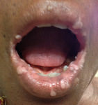
- Title:
- Condyloma Acuminata, Mouth, Image 3
- Description:
- Genital warts (or condylomata acuminata, venereal warts, anal warts and anogenital warts) are symptoms of a highly contagious sexually transmitted disease caused by some sub-types of human papillomavirus (HPV). It is spread through direct skin-to-skin contact during oral, genital, or anal sex with an infected partner. Warts are the most easily recognized symptom of genital HPV infection, and types 6 and 11 are responsible for 90% of genital warts cases.
- Keyword:
- Condylomata Acuminata, sexually transmitted disease, Genital wart
- Subject:
- DNA Virus Infections, Papillomavirus Infections, Virus Diseases, Skin and Connective Tissue Diseases, Skin Diseases, Skin Diseases, Viral, Sexually Transmitted Diseases, Tumor Virus Infections, Skin Diseases, Infectious, Sexually Transmitted Diseases, Viral, Condylomata Acuminata
- Creator:
- Metropolitan Hospital Center, Kathryn Russel, MD
- Publisher:
- Metropolitan Hospital Center
- Language:
- English
- Copyright Holder:
- Metropolitan Hospital Center
- Rights:
- http://www.i-human.com/service-agreement-print
- Resource Type:
- Photo

- Title:
- Condyloma Acuminata, Genitals, Image 2
- Description:
- Genital warts (or condylomata acuminata, venereal warts, anal warts and anogenital warts) are symptoms of a highly contagious sexually transmitted disease caused by some sub-types of human papillomavirus (HPV). It is spread through direct skin-to-skin contact during oral, genital, or anal sex with an infected partner. Warts are the most easily recognized symptom of genital HPV infection, and types 6 and 11 are responsible for 90% of genital warts cases.
- Keyword:
- sexually transmitted disease, Condylomata Acuminata, Genital wart
- Subject:
- Skin Diseases, Infectious, Virus Diseases, Skin Diseases, Viral, Sexually Transmitted Diseases, Viral, Tumor Virus Infections, Papillomavirus Infections, Condylomata Acuminata, Skin Diseases, DNA Virus Infections, Skin and Connective Tissue Diseases, Sexually Transmitted Diseases
- Creator:
- Metropolitan Hospital Center, Kathryn Russel, MD
- Publisher:
- Metropolitan Hospital Center
- Language:
- English
- Copyright Holder:
- Metropolitan Hospital Center
- Rights:
- http://www.i-human.com/service-agreement-print
- Resource Type:
- Photo

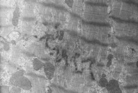
- Title:
- Heart, Cardiac Tissue Continuous Capillary, Transverse Section; Transverse Section, Homo
- Description:
- Electron micrograph and H&E
- Keyword:
- microscopic, EM, Heart, TEM, cardiac tissue, heart, Microscopy, Electron, Scanning Transmission
- Subject:
- Diagnostic Imaging, Cardiovascular System, Histocytological Preparation Techniques, Histological Techniques, Microscopy, Staining and Labeling
- Creator:
- John Cotter, PhD
Department of Pathology and Anatomical Sciences
University at Buffalo School of Medicine
- Publisher:
- University at Buffalo School of Medicine
- Language:
- English
- Copyright Holder:
- University at Buffalo School of Medicine
- Rights:
- http://www.i-human.com/service-agreement-print
- Resource Type:
- Slide
- Identifier:
- 2125
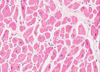
- Title:
- Heart, Cardiac Muscle Longitudinal Sections And Intercalated Disc
- Description:
- H&E and Electron micrograph
- Keyword:
- Heart, heart muscle, heart, TEM, EM, intercalated disc, Microscopy, Electron, Scanning Transmission, microscopic
- Subject:
- Staining and Labeling, Cardiovascular System, Histological Techniques, Diagnostic Imaging, Histocytological Preparation Techniques, Microscopy
- Creator:
- John Cotter, PhD
Department of Pathology and Anatomical Sciences
University at Buffalo School of Medicine
- Publisher:
- University at Buffalo School of Medicine
- Language:
- English
- Copyright Holder:
- University at Buffalo School of Medicine
- Rights:
- http://www.i-human.com/service-agreement-print
- Resource Type:
- Slide
- Identifier:
- 2126
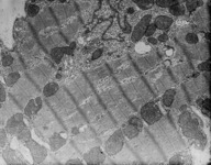
- Title:
- Heart, Cardiac Tissue Continuous Capillary, Transverse Section; Transverse Section, Homo
- Description:
- Electron micrograph and H&E
- Keyword:
- Heart, Microscopy, Electron, Scanning Transmission, microscopic, TEM, cardiac tissue, EM, heart
- Subject:
- Microscopy, Histocytological Preparation Techniques, Diagnostic Imaging, Histological Techniques, Staining and Labeling, Cardiovascular System
- Creator:
- John Cotter, PhD
Department of Pathology and Anatomical Sciences
University at Buffalo School of Medicine
- Publisher:
- University at Buffalo School of Medicine
- Language:
- English
- Copyright Holder:
- University at Buffalo School of Medicine
- Rights:
- http://www.i-human.com/service-agreement-print
- Resource Type:
- Slide
- Identifier:
- 2125
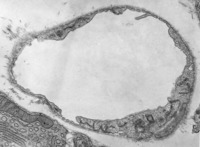
- Title:
- Capillary - Fenestrated And Thryoid Continuous
- Description:
- Electron micrograph
- Keyword:
- EM, TEM, thyroid continous, fenestrated, capillary, microscopic, Microscopy, Electron, Scanning Transmission
- Subject:
- Microvessels, Cardiovascular System, Capillaries, Blood Vessels, Microscopy, Diagnostic Imaging
- Creator:
- John Cotter, PhD
Department of Pathology and Anatomical Sciences
University at Buffalo School of Medicine
- Publisher:
- University at Buffalo School of Medicine
- Language:
- English
- Copyright Holder:
- University at Buffalo School of Medicine
- Rights:
- http://www.i-human.com/service-agreement-print
- Resource Type:
- Slide
- Identifier:
- 2124
- Title:
- Heart, Cardiac Muscle Longitudinal Sections And Intercalated Disc
- Description:
- H&E and Electron micrograph
- Keyword:
- microscopic, TEM, EM, Heart, heart, heart muscle, Microscopy, Electron, Scanning Transmission, intercalated disc
- Subject:
- Diagnostic Imaging, Microscopy, Staining and Labeling, Cardiovascular System, Histocytological Preparation Techniques, Histological Techniques
- Creator:
- John Cotter, PhD
Department of Pathology and Anatomical Sciences
University at Buffalo School of Medicine
- Publisher:
- University at Buffalo School of Medicine
- Language:
- English
- Copyright Holder:
- University at Buffalo School of Medicine
- Rights:
- http://www.i-human.com/service-agreement-print
- Resource Type:
- Slide
- Identifier:
- 2126
